Sunday, September 1, 2019

Final Remarks - CEC

Hello again,

I was very sad to be leaving the Cleveland Equine Clinic at the end of the summer. The veterinarians go above and beyond to teach students. If you are looking to experience a summer of ambulatory practice with a few hospital patients sprinkled throughout, I would HIGHLY recommend this experience. I was out on the road every day and rotated through riding with all of the vets at the practice, giving me diverse experiences as each vet has their niche within the practice. I lived on the practice grounds, allowing me to do physical exams and treatments on the hospital patients at the clinic. Here is a brief list of clinical work that I experienced this summer:


  • Standing MRI 
  • Caslicks surgeries
  • Castrations 
  • Physical Exams 
  • Lameness Work-ups 
  • Respiratory, lameness, and colic ultrasounds
  • Field necropsy 
  • Dental examinations, floats, and extractions
  • Blood draws and assessing blood work 
  • Foal handling
  • Reproductive exams, ultrasounds, and AI 
  • Endoscopic examinations
  • Gastroscopic examinations 
  • Radiographs 
  • Ophthalmology examinations 
I can't thank the veterinarians and staff enough for the time and effort they spent teaching me about veterinary medicine. I'm excited to be entering my final semester of the didactic curriculum and will utilize the knowledge that I gained this summer throughout my clinical rotations. 

Friday, August 2, 2019

Peterson & Smith Weeks 7-12!

Hello! Here's a final recap from Peterson and Smith and some suggestions if you are thinking about doing your fellowship here. I loved the opportunity to complete a fellowship through the Equine Fellows program and highly suggest the program to other students interested in pursuing Equine Medicine. Over my last six weeks I got to get into surgery, go out on the road, and spend some more time in the hospital as well. Peterson and Smith also have an Equine Reproduction Center. Having so many opportunities in one clinic is one of the things that made me want to spend my summer in Ocala!

Surgery
During my time in surgery I saw three arthroscopies, a hoof mass removal, and three emergency colic surgeries. I had a lot of fun getting to see these surgeries! This was my first time ever getting to go into surgery. Watching how they get the horse from incubation to surgery and then into recovery was really exciting. The emergency surgeries were my favorite. The rush of getting the horse to the table as quick and safely as possible to get the best outcome was really neat. The surgery techs are really fun to work with and they explain what is going on and what will happen next the whole time throughout the surgery. If it was slower I'd ask the techs if I could go watch the surgeries that are going on. I highly suggest asking to go in to watch as often as you can! You can learn a lot about anatomy and what the surgeons are doing, and when I was the only student in surgery they would quiz me on what they were doing.

Ambulatory
This was one of the highlights of my time down here! Going ambulatory gave me the opportunity to learn from vets first hand and ask lots of questions regarding our cases or being a vet in general. On my first day ambulatory I got to go on the road with Dr. Rood. It was HOT this day (like 108ยบ!!). We got to do a lot of preg checks, scan some foals for pneumonia, and microchip several foals! The foals travel between Kentucky and Florida so it is important to scan both sides of their necks to check for microchips before placing one. The next few ambulatory days I spent with Dr. Cadena. With Dr. Cadena I got to see a lot of preg checks, pull blood for coggins, see how artificial insemination works on a Quarter Horse Mare, and a few other fun stops. The best part about ambulatory is that our days were always so different. Another bonus was seeing some of the beautiful farms around here! It was my first time getting to travel around and see the countryside of Ocala.

Hospital
The recent excitement around here has definitely been colics! We have seen a lot of different types of colics over the last few weeks. I have gotten comfortable with the way that the doctors work up colics and how to set up for them. During my second to last week we saw a nephrosplenic entrapment and a displaced spleen in the same day, both were treated with Phenylephrine and then went for a jog up and down hills at the clinic.
Another case that we saw a lot were Pneumonias. Pneumonias were diagnosed with a combination of a transtracheal wash, an ultrasound, and radiographs. Rhodococcus equi pneumonia is extremely common in foals and something that is discussed often around the clinic. This type of bacteria lives in the soil and is one of the reasons why they suggest foals to be born in stalls in Florida. If one foal at a farm gets infected with this bacteria, it would be common to see others from the same farm present with pneumonia as well. We monitor for respiratory distress, respiratory effort, and rectal temperature in these foals as often as every 1 to 2 hours.
A fascinating piece of equipment I saw during the last few weeks in Ocala was the Lameness Locator. This device was able to be attached to the horse and the horse would be jogged, tracking his or her lameness on an iPad. Once the initial tests were completed, the horse then would have nerves blocked on the leg that it indicated. The program would put together charts that could be compared, showing the degree of improvement between the initial lameness and different diagnostic tests to assist in locating the issue.

Suggestions:
  • A good pair of shoes! I purchased these boots from Tack Shack of Ocala. They broke in really quickly, were extremely comfortable, and held up through everything over the summer. Having a good pair of boots was essential. It took me a week or so to find what I was looking for and definitely wish I would have purchased these immediately. I highly suggest finding a good pair before starting work because you do work hands on with tons of horses on Day 1.
  • Housing! The housing is extremely comfortable. Everything you could ever need is already down there for you. I chose to bring my own bedding, but bedding is provided if you would prefer that. I purchased a 5 drawer organizer for my clothes which was helpful to keep all of my items together.
  • Groceries - there is a Publix only ~5 minutes from the house! They had so many deals every week. Also every Thursday the clinic hosts Food Truck Thursday. The menu is posted at the beginning of the week in the break room!
  • Restaurants - our favorite restaurant was Sammy's! Sammy's is only a few minutes from the house and have amazing pizza. Las Margaritas and Yummy House were other favorites that the technicians at the clinic suggested that we loved. During one of my last weeks we found Ocala Downtown Diner - as a bonus it is open as soon as night shift ends for a really good breakfast.
  • Water. Water. Water. It is hot and going from Michigan to Florida is a huge change! I carried my water bottle with me everywhere!
  • Attend rounds! At 5pm every day the residents and interns walk through all of the barns to discuss each case and plans for that case over the next day or so. This is a great time to hear the updates on the patients and more details about how they are progressing. There is also Journal Club and Topic Rounds during the week that the fellow is invited to as well.
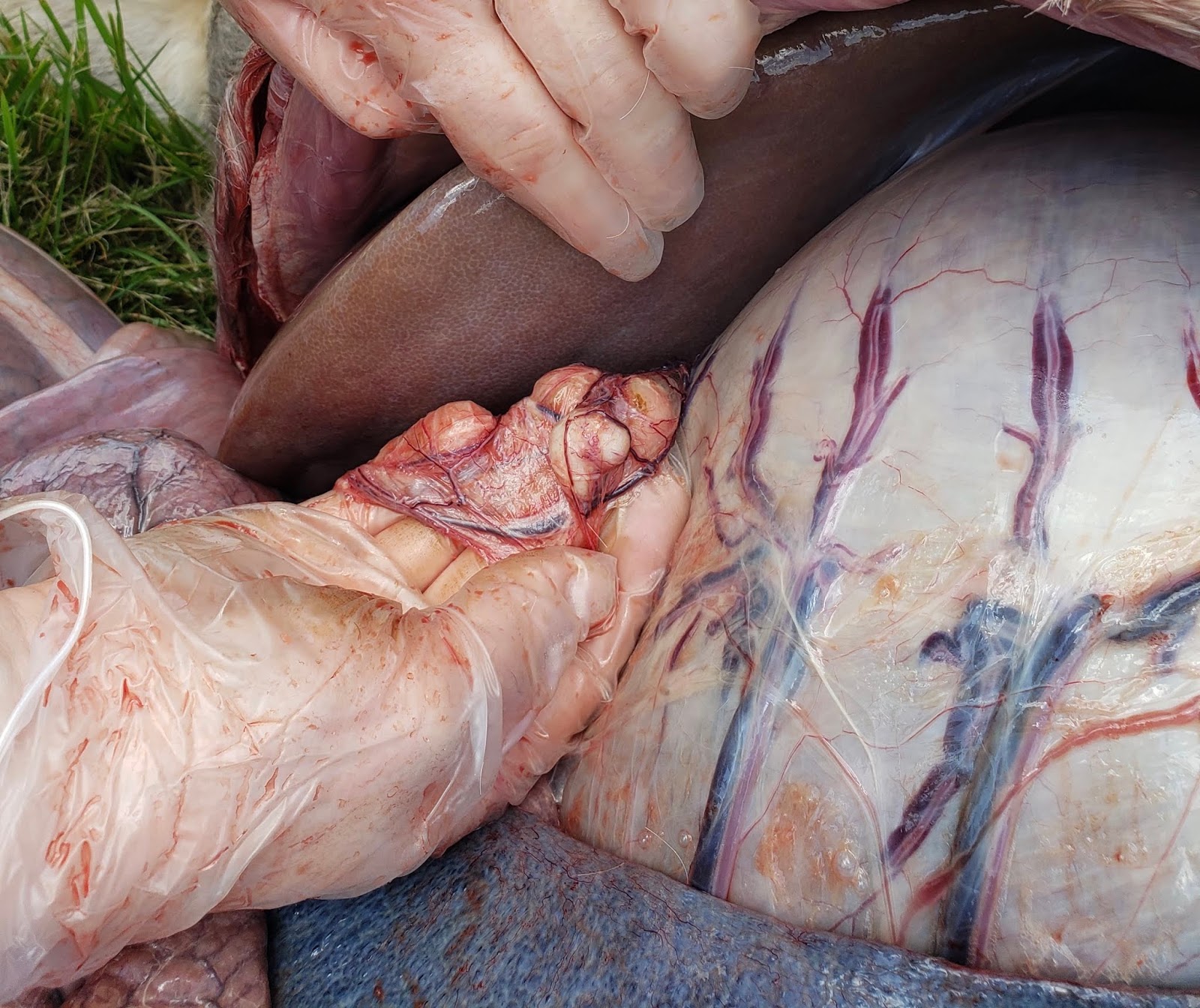
Overall, I had a great summer in Ocala and am so thankful for the experience Peterson & Smith and Michigan State offered me. I learned so much about how to care for foals and the preparation that went into getting horses into and out of surgery. Getting on the road for a few days was definitely one of the highlights of my experience. My favorite moment by far was when we had a mare foal out during one of my shifts. It was certainly unforgettable! I saw a lot of cases, got to watch a standing jugular thrombectomy and tendon sheath excision, and met a ton of amazing people.

Feel free to reach out if you have any questions about Peterson and Smith or Ocala in general. Thanks for reading :)

Tuesday, July 30, 2019

NJ Equine Clinic - Last Week!

We lost power this week and it was all hands on deck as we worked to get water to the horses not only in the hospital, but out in the fields as well. There was a crazy ~10 minute storm with high winds that caused many towns in NJ to lose power. Other than a limited supply through generator, our power was out from Monday night to Thursday afternoon— but that didn’t stop us from seeing appointments! 


We saw an epiglottic entrapment in which the aryepiglottic fold (a normal part of larynx anatomy) envelops the epiglottis, potentially leading to poor racing performance. The entrapment is diagnosed by endoscope and in this case the aryepiglottic fold was transected to free the epiglottis.


Case Highlight: EOTRH
Equine Odontoclastic Tooth Resorption and Hypercementosis (EOTRH) is a syndrome most commonly seen in older horses that results in resorptive lesions of the incisors and canine teeth.  EOTRH is typically diagnosed through radiographs but the pathophysiology of the disease is currently unknown. The only current treatment that exists is to extract the affected teeth.


Affected teeth are extracted due to tooth loosening and associated pain. Loose teeth can fracture off as in the case of this patient. Many horses actually do much better once the teeth have been extracted as they can go back to eating/being horses without feeling pain.


Thank you to everyone who read these posts and followed along during my time at New Jersey Equine Clinic!

A special thank you to the nurse staff at NJEC - Melissa, Alisha, and Danielle for letting us get in their way and sometimes take over, Dr. Leahy for her Spartan camaraderie and life lessons and lastly Dr. Smith for being ever so willing to answer questions and keep us on our toes while dishing out questions. I am so truly thankful for the experience and am forever grateful to have been taken in having had mostly small animal experience. Thank you for remembering what it was like to be a student and for giving me a chance in equine med. I can’t wait to share all of my acquired knowledge! 

Lastly, a million thank you's to Dr. Rashmir and Dr. Schott of MSU who without their support this invaluable experience would have never happened. 


Happy Summer Everyone! Off to our next adventure! 

Stefany Gomez 
MSU CVM 2021


Wednesday, July 24, 2019

A grab bag of cases at Littleton Equine

Hello all,
My time at Littleton Equine Medical Center is coming to a close. It's been a great summer learning in the ICU and meeting some amazing doctors, technicians, and fellow vet students from around the country. If you are an MSU CVM student thinking about applying for this position, please reach out to chat with me (denneyk1@msu.edu).
I thought I would wrap up my blog posts with a grab bag of unusual cases we have seen the last week or so.
In mid May a yearly Arabian presented to our emergency service for a hemoabdomen following a routine castration. Although I hadn't arrived at Littleton yet to watch this case work up, the doctors ran diagnostic blood work and gave the gelding a plasma transfusion. He did not improve enough with the transfusion, so the next day he was given whole blood and the clinical team started to investigate the reason for his hemorrhaging. Suspecting a possible coagulation issue, a coag panel was sent off. The results showed that this gelding had a defect in factor 8, a rare issue in horses. Factor 8 is part of the intrinsic pathway of the coagulation cascade, which meant his PTT time was elevated. He was diagnosed with hemophilia A. After the blood transfusion, the doctors were able to get his hemorrhage under control. The diagnosis was explained to the owners and he was sent home with instructions to watch him carefully and return to the clinic if he sustained any trauma because he would not be able to clot properly. This week our favorite little Arabian gelding returned with a hematoma the size of a basketball above his tuber sacrale. He had been frolicking in the pasture and took a spill into a T-post. The trauma caused him to hemorrhage and form the hematoma. He has remained in the clinic for close monitoring this week. The hematoma does not appear to be changing in size and he doesn't appear to be continually losing blood. We have closely monitored his mucous membranes, CRT, and heart rate on an hourly basis to assess if he is losing blood. The hemotoma was ultrasounded to confirm it was in fact blood filled. This current plan is to go home on stall rest until the hemotoma resolves and to remove as many dangerous items from his pasture as possible. Although our surgeon wasn't thrilled about the idea of having to take a hemophiliac horse to surgery, we discussed the safest ways to do that if it had to happen. One note our internal med doctor made was that vasopressin stimulates the release of more factor 8, which it is an expensive medication, but would be helpful to control bleeding in this case if surgery was necessary. Luckily, this guy doesn't currently need to go to surgery.

This summer we have also seen a few horses with severe ulcers. One of the most effective ways to treat them is with an SPL (sub palpebral lavage system). This is where a medication diffusing device is placed under the eyelid and a line is run up the horses mane. Most horses do not respond well to having eye medication placed daily, so an SPL helps to deliver medications in a safe and reliable manner. We usually give "ulcer mix" which has gentamicin, +/- atropine, artificial tears, and acetylcysteine. We also give serum via the SPL to help with the healing process. I had never seen an SPL before coming to Littleton, so it has been fascinating to watch the placement of them and how well they help to heal ulcers.

My last interesting case for this week was a mule who came in for a ovariohysterectomy (spay). This isn't commonly done in horses because their heat cycles are controlled well with hormonal medications like Regumate, but this mule had severe behavioral issues and the concern was that they were stemming from hormonal issues due to a possible ovarian tumor. On ultrasound the left ovary was considerably larger than the right and was anovulatory. The concern was that this mule had a granulosa cell tumor (GCT) producing testosterone in her ovaries that was leading to her behavioral issues. The ovaries were removed via laparoscopic surgery and we will wait to see if the behavioral issues resolve. One way to test for GCT's is with an AMH and testosterone test. Her's was negative, but our surgeon has had that happen in the past and send the ovary in for histopath and it was in fact a GCT. This was another unusual case that was very cool to watch unfold.

It has been a roller coaster of a summer here in CO. I am very grateful for this opportunity and for all I have learned and the amazing folks I have met. Please always feel free to reach out if you have any questions.

All the best,
Kaity

Monday, July 22, 2019

A Field Necropsy - CEC

Hello again! This past week we had a case that turned into an amazing learning opportunity. We were called out to evaluate a pony that had a history of founder. When we arrived, you could tell from a distance that the pony was having trouble breathing.

This is what we found on our physical exam:

Heart Rate: 80 beats per minute
Respiratory Rate: 100 breaths per minute
Mucous Membranes: Brick red with a toxic line
Capillary Refill Time: Prolonged
Digital Pulses: Within normal limits
Abdomen: Extremely distended

The pony did not act sore on his feet. When we lifted his feet, he was not reluctant to put weight on the opposite front foot. A lot of horses with founder would be resistant to this due to how painful their front feet can get. Additionally, he was not standing in a way that would shift weight off of his front feet. We were unable to do rectal palpation due to his size.

Unfortunately, due to the pony's poor condition and old age, the owner elected to humanely euthanize the pony. However, she agreed to let me ultrasound and then necropsy the pony for learning purposes. It was extremely kind for her to allow me to do this.

The ultrasound yielded some additional information. When scanning the lungs, we saw "comet tails." This suggests that there is some level of disturbance on the outside pleural surface of the lungs. Dr. Sarah Varnell was concerned that there may be fluid in the thoracic cavity as well because of the scan, which could have been contributing to his labored breathing. We did see some nodules on the spleen and there was decreased motility of the small intestines. We weren't able to do the best abdominal scan, because we only had a tendon ultrasound probe on the truck. But we did the best that we could with what we had!

It was great to get the opportunity to match the ultrasound findings to the necropsy findings. When we opened him up, immediately we noticed that the ventral colon was extremely distended. The vasculature of the intestines and stomach was also incredibly distended. The lymphatics of the mesentery were also so engorged that we initially thought that the abdomen had parasites in it! At the hilus of the mesentery, the blood vessels and lymphatics were so distended that when you reached your hand into the abdomen, they felt like ropes. There was indeed fluid sitting around the lungs and hemorrhagic fluid in the abdomen as well. The small intestines were abnormally colored. There were in fact nodules on the spleen and also on the kidney. The liver lobes were rounded. Although there was a lipoma, it was not of any concern for intestinal strangulation.

While we did not come up with a definitive diagnosis because no samples were sent off for histopathology, Dr. Varnell was suspicious of lymphoma given the pony's age and necropsy findings. This case was truly incredible. To get to work up a horse, perform ultrasound diagnostics, and then compare those findings to the necropsy is an experience I will never forget. I am very humbled to have had this opportunity and that the owner values student education so much.


Images:
Heart and Lungs

Spleen with nodules
Abnormally colored small intestine
Spleen with irregular consistency
Distended stomach vasculature with adjacent lipomas
Distended vasculature and lymphatics
Distended colon
Vessels that resembled "ropes"


Colic craziness at Littleton Equine

Hello all,
I hope summer is treating you well. It has been so busy here at the clinic lately. Our ICU is full and most of the cases are colics. I thought I'd give a brief overview of what colic means and how we address it. 
Horses colicing has been a very common occurrence this summer. Colic is a general term for any abdominal discomfort a horse experiences. Colic's can have many causes, ranging from an impaction of feed material, gas colic, sand colic, strangulation of the gut, entrapment of the gut, or even when the gut ruptures. To work up a colic we start with a basic physical exam including:  temperature, listening to gut sounds, check mucous membranes and CRT, feel digital pulse, and get a heart and respiratory rate. We will also get a history of feed and medications from the owner. Then we will then do a rectal exam on the horse to feel for dissented loops of small intestine, to feel if the colon is caught between the kidney and spleen (a nephrosplenic entrapment), or to see if we can feel an impaction in the cecum or colon. We then pass a nasogastric tube into the stomach to see if the horse will reflux. Each patient will also be ultrasounded to help assess what type of colic is occurring (can you see the kidney and spleen, are there dilated loops of SI, ect). We will also run a CBC, chem, and lactate on the patients. A very helpful piece of blood work is the lactate. Lactate is a byproduct of anaerobic metabolism, so if the lactate is high it indicates something is depleted of oxygen, and commonly with colics it is due to strangulated/dying gut. Depending on what we finds on the physical exam and with diagnostics, the patient is either determined to be a medical colic or a surgical colic. If the horse needs surgery, then they will head up to the clinic to our surgeons. If they are a colic that can be treated medically, we will give them NSAID for pain management, start then on a Lidocaine CRI for pain and to increase GI motility, IV fluids, and sedation. If the patient continues to be painful, we can add in additional medication like Buscopan, more intense pain meds, and more effective prokinetics. If the patients pain can not be controlled, then we either have to go to surgery or we have to euthanize. This week I got to see a colic surgery which was a large colon torsion. Unfortunately the torsion had cut off enough of the blood supply to the colon that is was too compromised and the prognosis for the patient was not good. We ended up euthanizing on the table. It was a sad case, but the owners felt that they had truly done everything they could for their horse and were very kind and thankful to us. One of our clinicians like to say that a little over half of colics with resolve on their own, 30% need some sort of meds and basic care, and around 15% need surgery. Working in the ICU we see so many really sick ponies that I have to keep in my mind those numbers because it is nice to know that many more horses are still out in the world happy and healthy. Please let me know if you have any questions (denneyk1@msu.edu). 

Thanks for following along :)

NJ Equine Clinic Week 5

This week we went on a farm call to see a horse with recurrent airway obstruction (RAO), or “heaves.” RAO is an inflammatory condition of the lower airways and is thought to be caused by inhaled antigens such as dust from food and bedding. Clinically, you could see coughing, exercise intolerance, increased respiratory rate and effort, flared nostrils and a double expiratory effort resulting in the characteristic “heaves line.” The most effective treatment for RAO is environmental change! 

Case Highlight: Rhodococcus equi
A foal presented with a swollen stifle, no fever, and no loud moist crackles upon listening to its lungs. This foal was currently being treated medically, but treatment did not appear to be working. If you look at the x-ray, you can see darker areas on the patella as well as the trochlear ridge of the femur, in this case indicating bone lysis. The U/S also revealed gas bubbles under the skin, another sign of bacterial infection. 


What could be the possible cause? Rhodococcus equi is a bacteria that is the most serious cause, although not the most common cause, of pneumonia in foals 1-6 months of age. Besides pneumonia, foals can also present with gastrointestinal issues and/or joint swelling. Rhodococcus equi is difficult to manage as it is ubiquitous in the soil and is also seen in the feces of horses and ruminants, thus providing vehicles for passive carriage. 
Our foal presented with joint swelling and thus was taken to surgery to have the stifle flushed and some of the loose bone around the patella and femur debrided. A sample was taken for culture.


Lots of birthdays this week! My mother, Tundra and I all got a little older (and hopefully wiser!) this week. Happy Birthday to Us! 


Hope you’re all having a lovely summer, time is flying!