Thursday, June 29, 2017

Weeks 1-2 at Littleton Equine Medical Center

I can’t believe how quickly the past few weeks (and the majority of the summer) have blown by so quickly!  I got a later start out here than I would have liked because I was in Washington DC doing the Smith-Kilborne project, which was also a great and insightful experience.  The first few weeks out here have involved a lot of ICU training as well as learning who the doctors, techs, and assistants are (there are 21 veterinarians including interns as well as techs and assistants for each), where everything is located, and learning the general routine here.  The majority of the work that I have done so far has been in the ICU, although my schedule is changing now to include days of observing and assisting with surgery as well as working with veterinarians here in the field.  One thing that I love is that every Monday morning, there is a staff meeting with case presentations and discussions of relevant current topics. 

The first few weeks in the ICU had some slow days, so I was able to go watch procedures and workups in the exam rooms in the main part of the clinic.  I feel like such a nerd being so excited about seeing diseases and conditions that we learned about in vet school, but I have gotten to see some really interesting cases lately!  My third day here, I came in partway through a procedure where they were doing a local block on a horse’s croup/hip area and oddly, started seeing bubbles coming out of the horse’s skin as the needle was removed.  As I heard the attending vets discussing the information about the presentation and history of the horse, such as that it had received an intramuscular injection in that location a few days prior, I realized that this horse had clostridial myositis and they were preparing to do surgical fenestrations in the skin at that location.

As a refresher for what clostridial myositis is…clostridial bacteria (commonly Clostridium perfringens type A, which is a Gram negative anaerobe) can either be inoculated or lie dormant in muscle tissue as spores.  They can convert to their vegetative, or active, form if there is sufficient trauma or irritation to their surrounding environment of skeletal muscle.  When that happens, they often release gas in the local tissues (so you may feel crepitus upon palpation) and can release some potent exotoxins that can potentially cause a systemic toxemia in the horse.  Although cases of clostridial myositis are frequently associated with intramuscular Banamine injections, they can also be caused by other intramuscular injections or even simple tissue trauma.

Fortunately, clostridial myositis is relatively uncommon.  Surgical fenestrations are necessary to perform because the bacteria must be exposed to oxygen in order to destroy them and combat the infection.  Prior to making incisions, the attending vets used ultrasonography to evaluate the extent of the infection so that they could determine where the cuts would need to be placed.  The underlying tissues were also debrided and the horse was later put in the ICU to be monitored and recover with IV potassium penicillin, supportive care, and daily wound cleaning and debridement.

The following day was another light day in the ICU so I came up to the clinic later in the day to observe and help with more cases.  A horse came in that had a high fever and was acting “off.”  He had been turned out with other horses, and had been bitten on the shoulder about 5 days prior to coming into the clinic.  The majority of his right side was uneven looking and he had ventral edema down his right side.  Again, crepitus could be palpated dorsally on this horse and after the shoulder wound was evaluated with cytology, clostridial myositis was again diagnosed.  The infection was more extensive in this horse and spanned from his shoulder to the end of his abdomen from about ¾ of the way up dorsally down to his ventrum.  Again, he was evaluated via ultrasound and was treated with surgical fenestrations, lavage, potassium penicillin, and also gentimicin because his white blood cell count was lower.  A major concern with treating horses with clostridial myositis is the potential complication of laminitis as a result of the systemic toxemia, so these horses were also placed in ice boots and Easy Rides (as well as having received general supportive care) and fortunately had no major complications.

I have also gotten to help with lameness and prepurchase exams both at the clinic and on farm calls and have had the opportunity to observe some interesting surgeries.  The most interesting one so far has been a carpal arthroscopy, not because they are very uncommon, but because it was the first time that I have been able to see an equine surgery that wasn’t performed as a standing procedure.  The arthroscopy was successful, and a large osteochondral fragment was removed from the horse’s carpus that had been lodged between the distal radius and the radiocarpal bone.  I learned how to recognize fibrillated cartilage and full thickness erosions, and Dr. Devine then finished the procedure by performing microfracture on the full thickness erosions to help stimulate fibrocartilage growth.

There have also been many, many colicky horses that have come in.  Two horses were also treated that came in with a rectal tear, but unfortunately both had to be euthanized despite great effort to save them.  That lead to a very useful discussion during the staff meeting on diagnosing the presence and extent of rectal tears and complications, a review of treatment methods, and good practices to help reduce the risk of causing one.

Overall, my first two weeks have been a blast and I am excited to continue learning from everyone here!  Everyone has been very kind and helpful, and I feel like I’m finally starting to get the hang of things around the clinic.  There has also been a fair bit of turnover lately because the old interns just finished up their year here and the four new interns have recently arrived.  Also, new externs arrive every two weeks, so that has been a good chance to meet and mingle with other vet students from other schools.  I have learned a tremendous amount in my first few weeks already and am looking forward to continuing to learn and contribute here for the rest of the summer!


-Calli

Saturday, June 24, 2017

Equine Athlete: Week 4

Show prep, show prep, show prep.  That was the theme of the week as we pushed to prepare horses for the two regional shows at the end of June.  Region 13 in Springfield, Ohio and Region 14 in Lexington, Kentucky.  Regional shows provide opportunities for horses to prove themselves on the road to the National shows coming at the end of the show season.  But, in order to get there, the horses must perform at their best throughout the Regional weeks.  So, we spent the last week traveling to many farms to prepare horses for both shows.

A day of show prep begins with a long list of horses.  We will fetch the horse and lunge them for the doctors to evaluate their movement.  Then, handle them as we work through flexion tests in order isolate the source of any lameness.  If locating the source is difficult we will go through and block certain regions and reevaluate.  Blocking a region of the leg is performed by essentially numbing the nerve supplying sensation to the intended area.  A lot of the blocks we perform are for the distal limb and foot.  Common blocks include the palmar digital or PD block that numbs the heel and sole of the foot.  Moving up the leg we can block the entire foot and back of the pastern with an abaxial sesamoid block.  As you move further up the leg you can block out more and more area in an attempt to find the source of the pain.  You want to start low and work up to prevent masking problems that are below the area that you block.  

Image 1. Dr. Hill injecting a hip
Once all of the horses are looked over, the doctors will sit down with the trainer and discuss treatment plans for those who need it.  That is when the real fun begins!  A lot of the treatments we do on farm are joint injections.  Intra-articular, or IA, injections go directly into the joint capsule to deliver the desired medication.  The most commonly injected joints are fetlocks, hocks, and stifles.  But it is not unheard of to inject the sacro-iliac joint (SI), hip, or shoulder.  This week we did a few SI joints and a hip (an injection I had never seen before) (Image 1).  Both the SI and hip injections are ultrasound guided to ensure that the medication is delivered to the desired location.

Image 2. Preparing to inject fetlocks
In order to inject a joint you must ensure that the region is extremely clean and that the horse is properly sedated to ensure a smooth procedure.  We begin by scrubbing the joint with a Chlorohexidine scrub that we alternate scrubbing and rinsing with a Chlorohexidine solution (Image 2).  We scrub and rinse until the area is properly cleaned and then let the final scrub sit until just before we are ready.  Although this process may sound tedious, it is a small and necessary sacrifice to prevent a possibly infection from occurring.  Not only can an infected joint cause soreness and increased lameness.  But, it will also cause irreparable damage to the joint and surrounding tissues.  Thus, we stress a proper scrub technique.

Once the area is prepared Dr. Hill or Dr. O'Cull will perform the injection.  There are many substances that can be injected, depending on the desired effect.  Corticosterioids such as Vetalog (triamcinolone) can aid in the reduction of inflammation within the joint.  Hylaronic Acid (HA) will be injected to supplement the HA naturally occurring in the joint and help increase lubrication.  Other injectibles include IRAP and antibiotics.  Most practitioners will use a combination of these substances, which is how Dr. Hill and Dr. O'Cull practice.  Studies have shown positive results for a variety of IA injectibles.  Therefore, as long as you maintain proper technique IA injections can prove to be extremely beneficial.

I can't wait to experience the excitement of Regionals and am excited to learn even more from these fantastic veterinarians.  I have already learned so much in the few short weeks that I have been with them.  Stay tuned for updates from the Regional shows and beyond.

Thursday, June 15, 2017

Equine Athlete: Weeks 2-3

My summer with Equine Athlete continues at a completely different pace than the one that was set at the busy Buckeye show during Week 1.  For the next few weeks we are scheduled to visit farms and evaluate horses of various levels and disciplines.  Both doctors are in and out of the state visiting farms around the country, while those of us at home are traversing Michigan and Ohio evaluating and treating as needed.  Most of the horses we are working with are slated to go to the regional shows coming up at the end of the month.  While others are less familiar clients that contact the practice for second opinions or more difficult lameness cases.  Although very different from Week 1, I am getting more time to learn the intricacies of lameness and how to treat different causes.  From young horses in training with training injuries, to seasoned show horses with chronic ailments, to solid children's horses with recurring unsoundnesses, we will be seeing it all over the coming weeks!

Image 1. Pre-purchase from 2015
Image 2. Radiograph from 2017
I have spent most of the past two weeks with Dr. O'Cull, as Dr. Hill has been traveling in and out to various farms around the country.  Our typical day consists of travel to different farms, evaluation of the patients, discussion with the client about treatment options and then performing treatments. Unlike the show cases, where we were treating in order to ensure top performance, these horses typically have more intricate diagnoses.  One of the more surprising cases was that of a gelding with suspected fetlock lameness.  Equine Athlete performed a clean pre-purchase on the horse only two years prior to him going lame during his training.  After performing a flexion test and isolating the source of lameness we opted to take radiographs of the region.  Immediately we identified the problem, advanced arthritis of the fetlock.

Osteoarthritis (OA) results from the progressive degeneration of articular cartilage.  This degeneration can have many causes; including trauma to the joint, progressive wear due to high impact activities, or abnormal/diseased cartilage.  Once the arthritic process has begun, there are no current therapies that can reverse the damage.  Luckily, there are options for slowing the progression of OA.  Limiting impact on the joint by decreasing workload, oral joint supplements, and intrarticular joint injections are some of the more common options for OA management.  Unfortunately for this horse, future top level performance options will most likely be limited due to the fast appearance and advanced stage of its OA.  The owners opted to inject the horses front fetlocks and place it on oral joint supplements.  We will continue to monitor the case and hopefully be successful in making the horse more comfortable and sound for many years to come.

What I am enjoying most about my experience with Equine Athlete thus far, besides the fantastic staff, is the opportunity to problem solve with an objective in mind.  All too often in medicine, we focus on simply 'making the animal better'; patching them up and sending them on their way.  But, what does that mean in the long run?  For the horses we are working with, it is straightforward.  Our job is to locate the source of their lameness and manage their physical well being for a long life of performance.  Then, when their career as a show horse is over, we are ensuring them a retirement into lighter work, lower level showing, or breeding (depending on the situation).  Not only are we helping their immediate ailments, but also providing for their long term soundness.  This is a way that I like to practice medicine and I look forward to my next adventures with Equine Athlete in the coming weeks.

Sunday, June 11, 2017

Rood and Riddle Weeks 1-4

Hello everyone! My name is Rachel Butler and I am completing my Equine Fellows at Rood and Riddle Equine Hospital in Lexington, KY for the summer. I am working as a surgery tech. It has been very busy at RREH and the time has flown by! I can't believe I am about to begin my 5th week! Over the past month, I have learned and experienced so much. The people I work with are wonderful and I am gaining many new friends.

As far as surgeries go, there are a lot of screws and joint arthroscopies, but I have also had the privilege of watching other surgeries, such as various fracture repairs, colics, tie backs, tie forwards, arytenoidectomys, tracheostomies, PTs, a pin cast placement, various eye procedures, abscess drains, joint flushes, and laceration repairs. I am sure there are a few more that I am leaving out. As a surgery tech, you basically have a front row seat to everything since you are in the surgery suite working.

I came in with very little experience with surgery and have really improved my knowledge and skills by working here. As a tech, your basic duties include dropping the horses for surgery, prepping the horses for surgery, work the surgeries, setting up for the next surgery, recovering the horses, and sterilizing instruments. While that doesn't sound like a lot as I write it out, there is a lot that goes into it, especially coupled on the case load of RREH. I have been in training for the past 4 weeks and feel as though I am just now starting to get the swing of everything. This past week I felt very accomplished as I was finally to the point where I could tech a case from start to finish without the observation or assistance of my supervisor! I hope to continue to build on my skills from here.

This weekend, I went to my parents' house in Columbus, OH to visit and ride my horse. The weekend has been very relaxing and I am getting ready to drive back to Lexington. I can't wait to see what this week brings. I will try to take some cool pictures this week for next week's blog entry. Happy Sunday everyone! Have a great week!


Saturday, June 10, 2017

MSU Large Animal Clinic


The first month has flown by and we have has some slow and some very busy days at the hospital. Each day brings some exciting cases, whether they end up staying for a week or just the day. In the past month we have seen a little bit of everything. Recently we had a Belgium mare come for dystocia. She presented in the afternoon, trying to actively push the foal out, this had been going on since about 8 AM that morning. The foal was deceased and after rectal palpation it was determined that they would not be able to pull the foal without anesthetizing the mare. After anesthetizing the mare they attempted to pull the foal, but with rigor mortis having set in they were unable to manipulate the legs and pull the foal out. A fetotomy was suggested. This was the first fetotomy I have seen. Although labor intensive, they only had to partially remove one leg before pulling the foal out. The mare recovered successfully and passed the placenta the next day before heading home. In the past month we have actually seen quite a few dystocias, mostly cows, but I have also assisted with one goat. Fortunately for the mare we did not have to perform a C-section, but most often that is what I have seen performed on the cows and goats.

Aside from daily hospital duties, routine lameness exams, and the occasional emergencies (sometimes not so occasional), I have also had the opportunity to help out with the Equine Theriogenology class. I have assisted with a couple castrations and one semen collection so far. Collecting from a stallion is pretty different than collecting from a bull, but it has been a great opportunity to be able to experience both.

Another interesting case that I have been lucky enough to help out with was a yearling that came in on emergency with anemia. The horse had a PCV of 12%, lactate of 18, and on ultrasound we found the blood swirling in the abdomen. With a highly suspecious diagnosis of hemoabdomen (a abdomenocentesis was never performed to confirm the diagnosis),  based on physical exam (heart rate and mentation), and PCV it was decided that the horse would need a blood transfusion. Luckily at MSU we almost always have universal blood donnor on site and we after some quick math, we were able to transfuse over five liters of blood. A heart rate, respiratory rate, and temperature have to be taken every five minutes when you are transfusing blood. The horse remained stable throughout the entire process and throughout the night. The next morning when I came into work the PCV/TS and lactate were re-evaluated and although the PCV remained fairly low (14%), the lactate decreased drastically from 18 mmol to ~1-2 mmol. The horse was much brighter in the morning and through the next coming days he would be almost back to his normal sassy yearling behavior.

Friday, June 2, 2017

Equine Athlete: Week 1

They say that trial by fire is the best way to learn.  After spending my first exciting week with Equine Athlete Veterinary Services, I can't help but agree.  I spent the first week of my internship in Columbus, Ohio at the Buckeye Sweepstakes Horse Show.  Ideally, I would have started a week earlier in order to familiarize myself with the equipment, protocols, doctors and technicians.   Unfortunately,  I was in Ireland for a few weeks and was therefore unable to start until the day that we left for the show.  But, learning on the fly has thankfully worked for me!

Before working with Equine Athlete I had little experience with the Arabian/Sport Horse show industry.  So, not only was this week eyeopening from a clinical standpoint but from an educational standpoint as well.  When we had a little downtime between patients we were able to watch some of the classes going on in the main ring.  After coming from a background in hunter-jumpers it was so neat to get to see these Arabian horses doing their thing.  One of the challenges I will face this summer is getting used to watching horses with more exaggerated movements for soundness when I am used to the less pronounced movements of hunter horses.  There is a lot to be learned from this fantastic practice and I am excited to have made it through Week 1.

Our whirlwind week at the Buckeye started when we got into town on Monday night.  We stopped into the fairgrounds to touch base with the trainers that had arrived already and get the list of the horses that needed to be checked out.  Thankfully all of the horses had shipped well thus far so we only had a few to do lameness evaluations on.  Until this point I had only been involved in lameness cases that we severe.  The horses we evaluate with Equine Athlete, especially at shows, are high level athletes.  This means that, more often than not, their unsoundness is subtle.  Our job is to pinpoint the locations of their soreness and help alleviate any pain.  That way they are able to perform at the top of their game.

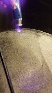
Image 1. performing laser therapy on
a sacroiliac region
At shows, your treatment options are limited due to USEF regulations on when certain procedures can be performed and medications administered.  Therefore we typically use a combination of chiropractic adjustment, shock wave treatments, and laser therapy.  Both shock wave and laser therapy were new technologies to me.  Shock wave therapy utilizes high energy acoustic waves to both dull pain and promote healing in injured areas.  Laser therapy uses high energy light waves to initiate healing processes in painful areas.  Both technologies can be implemented over many areas of the body if needed.  Some of the most common areas that we treated during the show were suspensory ligaments, thoracic and lumbar regions of the back, as well as sacroiliac regions of the pelvic area.  Much like treating sore muscles with heat or ice, these practices helped to ease the soreness and improve performance.

Alongside lameness cases we were the designated show veterinarian.  That means that any horses that get sick or injured during the show get treated by us.  Thankfully there were not many cases of sick horses during the Buckeye.  We did see a few colic cases and one laceration during the week.  All of these ailments were quickly corrected by Dr. Hill or Dr. O'Cull.

One of the most interesting cases throughout the week involved a young horse with unusual edema in the heart girth area.  The horse had pulled a shoe in its stall the day before.  The suspicion is that the horse got cast in its stall and strained its pectoral muscles in the process.  We performed an ultrasound of the area to determine if the swelling was subcutaneous or intramuscular.  After determining that the edema was intramuscular it confirmed the suspicion of a strained muscle.  The doctors prescribed dexamethasone and Equioxx to help relieve the inflammation.  The horse also received laser treatment to the area interspersed with icing and liniment application.  A few days of treatment resulted in almost complete correction of the swelling.  The horse was even able to show during the last few days of the Buckeye.

My first week was a whirlwind.  Between learning the ropes of horse show practice and the procedures and protocols that Equine Athlete practices I was tuckered out by the end of the week.  Thankfully the doctors and technicians were extremely helpful and were always willing to answer my questions.  I am enjoying the fast paced work and cannot wait to see what the rest of the summer has in store.