Sunday, June 30, 2019

New Jersey Equine Clinic Week 2

Here are some of the medical cases we saw this week:


Pituitary Pars Intermedia Dysfunction (PPID) + Maggots!
PPID is also known as Equine Cushing’s Disease. It involves an overgrowth of a portion of the pituitary gland (pars intermedia!) which leads to an excess production of hormones. PPID is most commonly seen in older horses and has been associated with a gradual onset of laminitis in about half of all cases. Repeated infections such as sole abscesses have additionally been associated with PPID, thus, enter the maggots. Medical-grade (don’t try this at home!) maggots were placed in pockets of the hoof where they are meant to chomp away at dead and infected tissues, while leaving the healthy tissue alone. The maggots will be allowed to feast for a couple of days, kept in place through the use of bandage material.

Choke + NG tube
Choke is the term used to describe an esophageal obstruction, usually caused by food. When the cause is food, the most common culprits are food that is too dry or coarse and food that swells in size once moistened ex. sugar beet. Often you will see saliva and feed material coming out of the horse’s nose and/or mouth. A scope was passed through one nostril to identify the location of the feed while a nasogastric (NG) tube was then passed through the other nostril in order to try and flush the material out. Water was pumped through the tube into the esophagus and then a mixture of gravity and pressure forces were used to allow the material to flow back out of the horse.


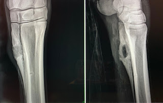
Case Highlight: Splint bone osteolysis
A horse presented to us with a swelling around the area of it’s splint bone. Radiographs were taken and they revealed osteolysis within one of the splint bones. The suspected cause was some sort of trauma that could have led to an infection. The horse was taken to surgery, and the area was debrided. 


The splint bones deserve more credit than at first given to them! When dealing with injuries to the splint bones, you have to remember that the heads of the splint bones come into contact with the horse’s wrists and hocks and thus help stabilize those structures. In addition, callus formation on the splint’s can lead to impingement on the suspensory ligament. 

It’s getting hot and humid here in NJ! Summer and relentless fly’s are upon us. Until next week!

- Stefany G

Saturday, June 29, 2019

Littleton Equine Week 5! Slithering Along- Snake Bites in Horses

Hello again from beautiful Colorado!
It has been very, very warm this last week in CO. Summer is officially here with temperatures in the 80's and 90's. The other two ICU summer students and I try to get out every weekend and explore a bit of CO. This weekend I think it will involve some sort of swimming to cool off. This last week has been great,  lots and lots of colic surgeries, a sinus flap surgery, a few sick foals, and a snake bit case. I haven't learned much about snake bits and how to manage them in vet school, so I am going to share this case and the medical treatments I learned because it was very intriguing to me.
To properly treat snake bites you have to be aware of what venomous snakes are in your local area. Here in CO there are mostly prairie rattlesnakes. At one of our morning meetings I got to speak with a couple of very seasoned field vets who know the CO landscape well. Their insight into where common snake bites are was amazing and really inspired me to get to know the geography and native animals where ever I will be practicing in the future. For example, usually the clinic sees 2-3 snakebites every summer, but some summers they see 10 snakebite cases from just one horse farm. There are quite a few management items that the vets recommend to those farms to help curb the number of bites. The snakes typically like rocky areas, so the vets recommend fencing those type of areas off. One of the older and wise vets even mentioned that pigs are able to eat the venomous snakes because they can survive the bites due to their increased body fat percent. So they have recommended adding a few pigs to the fenced in areas as a short term solution.
Horses are usually bitten on the muzzle, occasionally on the distal limbs, and rarely on their trunk. The muzzle is the most common bite site because they are down grazing and a snake bites them or they see one and go up to investigate it. Sometimes horses step on them and get bite on the distal limbs. One of the vets advised us that if a horse is showing clinical signs of a snake bite, but you can't find the fang marks in the normal locations, it could be because the horse rolled on the snake and was bitten its dorsal spinal area. Usually you will see two bloody fang marks where the snake bite the horse. One of the vets reported seeing a case one time that had bloody fang marks, but the horse didn't have any clinical signs of a snake bite. They contributed this to what is called a "dry bite", where the snake is very young or very sick and doesn't have venom to inject into the horse. The vet recommended still treating the horse with the typical treatment just to be cautious.
The common clinical sign of a snake bite is a  massive amounts of swelling in the area that was bitten. Bites to the face are much more severe because they have a greater blood supply to spread the venom. One of the most common issues with a bite to the face is that the swelling mechanically occludes the horses nostrils. A very simple trick to be able to help the horse breath is to insert 1 small section of garden nose into each nostril. This will provide the horse with a patent airway until the swelling can be managed.
The basic treatment for snake bites is to give the horse a steroid, such as dexamethasone, to decrease the swelling and inflammatory reaction. An NSAID, like banamine, can also be helpful to decrease swelling and control pain (NSAID's and steroids should not be given together though, so one or the other). Snake fangs have also been reported to carry bacteria like clostridium, so an effective antibiotic is also a vital part of treatment. Commonly horses that are bitten go into shock, so IV fluids are important to counteract that. If the bite is on a distal limb, a standing wrap and cold hosing can help to decrease swelling. If the bite is on the muzzle, fenestration of the muzzle with a 14 g needle can help to decrease swelling. The area that was bitten should be clipped and cleaned.
The latest development in treating equine snake bites is antivenom. From what the vets explained to us about snake bites, one of the long term side effects can be cardiac damage. This damage can be accessed by measuring troponin, a cardiac muscle enzyme, level right after the bite and a few weeks post treatment. One of the advantages of antivenom is that it might help to decrease the chances of cardiac damage because it neutralizes the toxins. Antivenom is very expensive for equine patients, but if the owner is financially able to afford the treatment, it is recommended. When administering the antivenom, the horse should be carefully monitored for signs of an anaphylatic reaction.
The case I saw managed here was given steroids, IV fluids, broad spectrum antibiotics, and antivenom. Cold hosing and stack wraps were also applied because the bite was on the LF distal limb. The clinicians also emphasized knowing important structures around the bite site because if a joint or tendon sheath is involved that would require more intensive treatment.
The venomous snakes here in CO do not produce a neurotoxin, but snakes in other areas of the country do and that specific envenomation requires a specific treatment.
The biggest lessons I learned about snake bite management in equine patients is that it is a medical emergency that needs to be treated quickly and effectively. Another big take away for me was to know the venomous snakes in your practice area because that will impact your treatment strategy.
Thanks again for reading along and please let me know if you have any questions (denneyk1@msu.edu). I continue to have a great time here in CO and will be back next week with more cool cases.

Best,
Kaity

Wednesday, June 26, 2019

Rood and Riddle Equine Hospital


Hey guys! Just a little update on my summer, sorry I haven’t been very active on here, I have just been crazy busy. Time is literally flying by, I cannot believe it is almost July. Working here at Rood and Riddle has had its ups and downs. Even though I’ve been here since May I am still learning new things every day, so I’m not as efficient as the other technicians but I’m getting there. I have learned a lot about technical things such as instruments, machines, and suture material which will be helpful for clinics and the rest of my career.

I am now consistently taking cases as a technician, which is very stressful because I don’t want to upset the surgeons. Like I said before there is a lot to know and every doctor does things differently. Since I am new to all of this I mainly help with transphyseal screws, arthroscopies, and colics. The more advanced surgery technicians are in charge of the more complicated surgeries such as fracture repairs and arthrodesis. Therefore I’m seeing similar surgeries every day, which to me is great, I am getting more and more comfortable with them each day.

With that, I am going to go through what a transphyseal screw (TPS) placement surgery looks like. First we start by prepping the leg, every surgeon is different on how they want it prepped. For example some have you clip with a 40 blade as others prefer a 10 blade. Prepping the patient for this surgery includes clipping, a rough prep with chlorhexidine and alcohol followed by a sterile prep. The prepping takes places in what they call the transfer area which is right outside of the operating room. Once you are finished prepping you will roll the horse into the room and hoist the leg and position the light on the surgical site. The surgeon will then drape and begin the surgery. He starts by making a small incision and then uses a drill bit to create a hole, next the a drill bit is inserted through the drill guide and the drill bit is advanced through the cortex. Most often 3.5 mm screws are placed in these foals. Once the screw is placed, digital radiographs are taken to insure that the screw was placed in the correct location. The incisions are then closed and bandages are placed. As a technician you are then responsible to take the horse to the recovery stall and page for a recovery person to take over your patient.

The purpose of the TPS placement is to correct a foal with angular limb deformity. This condition if left alone can cause lameness and affect a horses performance. Therefore, for sales purposes excellent confirmation is needed.

Let me know if you have any questions! I am still enjoying beautiful Lexington and don’t want to leave anytime soon. J

Tuesday, June 25, 2019

A Pony's Tale - Cleveland Equine Clinic

My favorite case of the week involves a schooling pony at a local farm. He came in from the pasture one morning so lame that he could barely walk. We did a full lameness work up which typically involves palpating the joints and tendons, evaluating movement, and taking radiographs. On palpation, there was a lot of pressure in the middle joint of the knee. My mentor suspected a fracture due to how lame the pony was at the time. We took radiographs and to our surprise, there was no fracture to be seen! 

Our next step was to tap the joint and see what the joint fluid looked like. Normal joint fluid should be clear and colorless. This pony's joint fluid was opaque and dark brown/red in color. We collected the fluid so that we could analyze it back at the clinic and injected an antibiotic, called Amikacin, directly into the joint. The owners of the pony were unable to provide the care needed due to the extent of the infection in the joint. However, the clinic owner and owner of the horse have a great relationship. We took the pony on as a charity case and brought him back to the clinic for treatment! 

Upon joint fluid analysis, there was a high white cell count, indicating that there was indeed an infection in the joint just as we had suspected. We decided to do an alternating schedule of procedures on the knee. The first night, the intern and I flushed the joint and then performed a regional limb perfusion. This regional limb perfusion involved putting a tourniquet on the leg in two places, one above the knee and one below. We then gave a high concentration of antibiotics into the vein near the knee. We left the tourniquets on for several minutes so that the antibiotics could absorb into the surrounding tissues and joint. This provides a strong and local source of antibiotics to help with the infection. It was a great learning experience for us both. The pony stayed in the hospital for about a week. We alternated flushing the joint and performing regional limb perfusions daily. 

Additionally, he was on Banamine for pain relief and systemic antibiotics. We took more radiographs after a couple of days in the hospital to make sure that there weren't any significant bony changes that can occur due to the infection in the knee. In a few short days, the lameness had significantly improved and the joint fluid was looking more like it should. At the end of the week, he was discharged and sent back to his farm to greet the next group of camp kids. It was truly an amazing chance to learn how to do regional limb perfusions and joint flushes. And best of all, there was a happy ending for the pony! This is just one of the many examples of how this clinic truly cares about student learning. Until next time! 

Liz Ritchie 

Sunday, June 23, 2019

New Jersey Equine Clinic Week 1

Hello from Millstone Township, NJ! My name is Stefany and I will be a third year veterinary student this upcoming fall. This summer my fellowship is taking place at New Jersey Equine Clinic, a referral hospital located in central NJ. Being a northern NJ native, it’s nice to be somewhat close to home and be able to spend some time with my family. While there is housing accommodation at the hospital for students, I am actually staying in a beach town about thirty-five minutes away with my boyfriend Bryan and our dog Tundra. I have to take full advantage of the ocean while I can!

The hospital owner and chief of surgery is Dr. Jen Smith. A recent hire to the team is associate Dr. Liz Leahy, an MSU grad. GO GREEN! Dr. Leahy completed her intern year with Dr. Smith and the pair have actually known each other for some time as Dr. Leahy started off as farm and stable staff at this very hospital while in college. 

As NJ Equine Clinic is a referral hospital, the majority of my first week was spent in the surgery suite. Some surgeries performed included: 

  • Bilateral cryptorchid - cryptorchidism refers to the condition in which one or both (in this case!) testicles do not descend normally into the scrotum. Testes start off in the abdomen in a fetus and need to make their way down past the inguinal canal into the scrotum. Sometimes they don’t make it all the way and need to be surgically found and removed. 

  • OCD Surgery - OCD stands for Osteochondritis dissecans and is a relatively common developmental disease typically diagnosed in young horses once they are worked. Malformed cartilage leads to the development of cartilage and bone flaps that can cause inflammation of joints and eventually lead to arthritis, so the “flaps” need to be removed. 

  • “Tie-back" procedure - this is one method used to treat laryngeal hemiplegia in horses, perhaps better known as “Roarers.” The noise made by these horses is due to paralysis of one or both arytenoid cartilages, cartilages of the larynx, due to lack of innervation to the muscle in charge of movement of the arytenoids on either side, respectively. 

Case Highlight: Arthroscopic flush of a navicular bursa 
This case became an emergency surgery as a horse was brought in that had stepped on a nail within a relatively longer time frame. With nail penetration into the foot, we primarily worry about compromise to the coffin joint, navicular bursa, and the tendon sheath. The fluid sample taken from the navicular bursa showed an elevated white blood cell count, indicative of an infection. During surgery, the bursa was flushed extensively (the solution to pollution is dilution!) and a regional limb perfusion was performed as well. See below for images!


Until next time! Here’s a photo of me with Danielle (LMU ’20) and Taylor (VT ’21), two students currently on externships. This was Taylor’s last week at the hospital, best of luck on your anesthesia rotation!


Friday, June 21, 2019

Littleton Equine Week 4

Hello all,
I am wrapping up my 4th week here at Littleton. Last night the clinic hosted the "In and Out BBQ". We just out a group of 4 new interns who are coming in and our 4 old interns are going out. The BBQ was a lot of fun and yummy food to say goodbye to our old interns and hello to our new interns. I had a good time, but left a bit early to catch some zzz's because I have a hard time transitioning from night shifts to day shifts. I spend this week in the ICU and got to spend one day with our surgeon, Dr. Hill.
I thought first I would tell you all about an interesting neuro case that presented last week and went home this week. A quarterhorse gelding came to the clinic for unknown neuro signs. He was ataxic, unstable at a walk,  had a lip droop, and had a head tilt. He eventually developed nystagmus. He reminded me of a dog with an inner ear infection. He was up to date on vaccines and there wasn't any history of trauma, but he did have history of cribbing. Cribbing is a bad habit where a horse chews on the wood in their stall or pasture.
All of the geldings bloodwork and physical exam perimeters (except for the neuro signs) came back within normal limits. The doctors on the case choose to shoot a few radiographs of his skull. The radiographs revealed bony proliferation around the temporohyoid joint. This lead them to the conclusion that this gelding had temporohyoid osteoarthropathy (THO). The cause of the THO was unknown, but it was a possibility that the cribbing lead to arthritis and damage of that joint. His clinical signs were due to the proximity of cranial nerves 7 (facial) and 8 (vestibulocochlear) to the damaged area. The bony proliferation was leading to nerve damage. The damage to CN 7 lead to the lip droop. The doctors also watched his eye on the affected side closely because CN 7 is also responsible for eye lid movement. If it is damaged it can commonly lead to ocular ulcers.  His other signs (nystagmus, ataxia, and head tilt) were attributable to CN 8. He was started on anti-inflammatory drugs and antibiotics. The antibiotics were in case the cause was bacterial in origin, which has been reported in some THO cases. When the gelding became more stable the doctors decided to do an endoscopic exam on him. The endoscope confirmed the THO diagnosis. After a conversation with the owner it was decided surgery was the gelding's best option.
The surgical option to treat THO is a ceratohyoidectomy. This procedure is done under general anesthesia and the goal is to remove the ceratohyoid bone, one of the bone that articulates with the hyoid apparatus. This helps to decrease pressure on the damaged joint and therefore decrease the pressure on the nerves. The surgery was successful with this gelding. He had mild clinical signs after the surgery, but they were slowly resolving. He went home this week and will continue to have routine rechecks to make sure he is making appropriate progress. It was a very interesting case to observe and a very happy ending for one of the sweetest geldings I've met.

Thanks for reading :)
Please let me know if you have any questions. Until next time!

Best,
Kaity

Sunday, June 16, 2019

Peterson and Smith Weeks 4-6

Hello again from Ocala! It's hard to believe I am already on my 6th week down here and 50% through the fellowship already. Over the past few weeks the clinic has entered the "slow season" according to the technicians. The case load has dropped a little and my posts will probably be more spread out from here on out. It appears that we may be done with having several foals in the clinic now and have migrated on to a surplus of colics (my favorite!).

Some highlights of the last few weeks are as follows:

  • Watching Dr. Kent perform an enucleation! This was the first enucleation I've seen and thought it was really cool! The eye was abscessed throughout so the doctor had to take a different approach to removing it. After it was out, she cut into it to examine the eye and see the extent of the abscess. This surgery was done in one of the treatment room's stocks so it generated quite a crowd and the other working students and I found it really interesting.
  • Foal presented for colic at 12 hours old. She passed meconium and normal manure, was nursing fine and despite about 10 minutes/hour of colic episodes, was an active and normal foal. After two days of intermittent colic episodes, the foal began resting quietly and was soon discharged. I was on night shift so I did not get to hear the full story on the foal, but I don't think they were able to get a definitive diagnosis. It felt like a puzzle and generated a lot of questions surrounding the case.
  • One 48 hour span of almost filling up Clinic Stalls where all of the colics go. During this time, the clinic had two mares (one with a foal, one in foal) go to emergency colic surgery, a gelding that presented with a mild colic while the doctors were in surgery, and more that presented with other medical types of colic. I think my shift saw 4 or 5 come in on emergency in two days!
  • A hawk that was hit by a car came to the clinic! A technician found him and we got to take care of the hawk for two days before he was able to be taken to a wildlife rehabilitation center. They took radiographs of him during the day shift and on nights we fed the hawk until he was able to be moved. I did not realize how big they were! The beaks were huge and it was an interesting change from seeing only horses for the last 5.5 weeks.
Having so many different horses in different life stages has been fun. I think my favorite part of this fellowship is seeing different horses from newborn foals to twenty-some year old retired Thoroughbreds. Growing up around Thoroughbreds definitely prepared me for this position. The one take away from the different age groups and how they present to the clinic is to make sure that you always treat the horse like you have not been working with it previously. When they settle into the clinic they may be the same or very different animals then they were when you worked with them the day prior.


At the end of this week I get to go home for a quick 4 day visit before coming back to begin my surgery rotation! I am really excited to be able to get into the operating room and see some exciting things. During our surgery rotation we are on call 24/7 in order to get the most surgery experience as possible. Unlike our day and night rotations, surgery is only one week so I will try and soak in as much as possible during my time there. When the surgery rotation is over, I am excited to be able to go ambulatory for a little bit of July and experience what it is like to be on the road.

Here's a bonus picture of the clinic's cutest neighbor :)


Thursday, June 13, 2019

Weeks 2-3 at Littleton Equine Medical Center!

Hello!
I am in my third week here in Littleton. The summer is already flying by. Most of my time has been spent in the ICU so far. I will usually work 3 days in the ICU and then do one day of surgery and one day in the field with an ambulatory doctor. In the ICU I am responsible for cleaning, lots of cleaning, stocking, doing treatments like feeding, handwalking, icing, giving IV and oral meds, and anything else the patient needs. I also get to assist doctors with whatever they need and get to see a lot of ultrasounds, x-rays, abdominocentesis, and general discussion about cases. I feel like I am really getting the hang of things here, which is wonderful because I feel like I’m helping more and learning more.

I think the most efficient way for me to blog is to discuss one interesting case from each week so things don’t get repetitive. I will discuss the case and then some general differentials and details about that general category of disease.
Today was a field day for me, meaning I got to ride around with one of our awesome vets and their tech all over the Colorado countryside. A lot of what we did today was dental work. Believe it or not, horses need routine dental care, just like you and I. It keeps them healthy, happy, and performing at their best. Performing dental work in horses is called "floating their teeth". It involves a good dose of sedation, usually Dormosedan, which is an alpha 2 antagonist that takes about 5 minutes to take action. Before the horse is sedated, a physical exam is done on them to make sure they are in good overall health and can safely be sedated. We listen to their heart, lungs, gut sounds (borborygmi), take a temperature, evaluate their overall weight and attitude, and ask the owner about any issues. After they are sedated, we place a oral speculum in their mouth. The speculum allows the doctor to get a better view of their oral cavity, but it also keeps the doctors hands and fingers safe from possible bite injuries. The doctor also has a head lamp for good lighting and a hand mirror to see all of the occulsal surfaces. The horses head is either held by an assistant or placed on a stand. The doctor can then start the float.
Tools for a float include mostly different size, textures, and shaped files. The files are used to smooth down rough points of the horses teeth, which I will explain more shortly. Some doctors prefer hand files, while others prefer power files that are attached to drills or specially made power tools (google power float for more details:). The important thing to remember with power tools used for floats is that their needs to be adequate irrigation to prevent too much heat and thermal damage, along with a vacuum system to remove debris that could be harmful to human and equine respiratory systems.
The whole reason a horses teeth need to be floated is due to their anatomy and eating habits. Horses teeth are different than human because they continue to grow all of their lives. Horse also chew their feed in a lateral motion and their upper arcade of teeth are wider then their lower arcade. The combination of these factors lead to "points" on the horses teeth that need to be rasped down. If they aren't filed down, they can create ulcers in the mouth that are very painful, they can limit the horses ability to eat, and they can impact performance because it is painful for the horse to have a bit in. Other issues can arise in a horses mouth that need to be addressed also. Some horses have poor confirmation that leads to unusual wear that also needs to be filed and corrected. A horse usually needs its teeth floated 1 time per year, but that can vary depending on the horse and circumstances.
Occasionally horses will need teeth extracted. First the doctor will do an oral exam and then take radiographs to access the health of the periodontal bone and root of the tooth. If they tooth is intact enough, it can be extracted orally. If the tooth is too diseased to safely be extracted orally, a tooth can be removed via the maxillary sinus. This morning we were able to do a recheck on a horse that had had this surgery done. It was a fascinating process to see.
Routine dental care for our equine patients is vital to keep them performing and feeling their very best. I have loved getting to see and help with floats. Please let me know if you have any questions (denneyk1@msu.edu). Stay tuned for more cases and interesting procedures :)

Best,
Kaity

Monday, June 10, 2019

Equine Athlete Weeks 1-4

Hi everyone! My name is Natalie Hershenson and I just completed my first year! This summer I am working for Equine Athlete with Dr. Brad Hill and Dr. Bailey Smith. Equine Athlete is an ambulatory veterinary service. We primarily service horses and barns that are involved with the Arabian horse show circuit. However, we are not limited to just Arabians – we see a lot of Saddlebreds and Quarter horses as well. Our primary job is a little confusing for those that are unfamiliar with the horse show world, so I’ll do my best to give the reader’s digest version of what we do. Dr. Hill has known most of these horses from the day they were born and has relationships with trainers and clients that dates back 10+ years. Every week or two before the big shows, we will go to the barns and do lameness exams and treatments on the show horses. These treatments are predominantly injections of the joints. Then we go to the shows that the horses/barns will be competing at and do additional treatments such as shock wave therapy, laser therapy, therapeutic ultrasound, chiro, etc. Our job, especially when pertaining to these show horses, is to help them perform as well as they possibly can at horse shows.

My first day we jumped straight into the deep end. We drove down to Ohio to one of the largest barns I have personally ever seen. The facility has over 200 horses in training and multiple trainers. The first day we do all the lameness exams – for over thirty horses. Lameness exams involve lunging the horse followed by flexions. This barn also rode every horse as well because sometimes specific problems only occur or are exacerbated under saddle. Needless to say, these lameness evaluations took us a full day. The next day we came up with a plan and started treatments. As stated before, these treatments are primarily injections. We don’t typically do injections at the horse shows because the horse needs three days off afterward. We also note if the horse needs to have therapy done at the show – which will offer a more immediate but short-term effect in comparison to injections. We then drove to Indiana to do it all over again at another barn. Week one was full of a lot of learning. I had to get my sea legs, as anyone does who starts a new job. I had to learn where everything was on the truck, learn how all the equipment works, what it’s used for, how to use it, how to prepare for treatments, etc. It was A LOT but Dr. Hill, Dr. Smith, and Christina were super patient and helpful. 

The second week marks the start of my first horse show. This horse show was held in Columbus, Ohio. We arrived on Monday and we stayed through the following Monday. This was a fun week for me. We were a tad slower than we usually are at horse shows, but this allowed me the opportunity to watch the classes. It gave me a chance to figure out what the heck was going on – what the significance of it all is, what our goal is, what we want the horse to look like, what makes a winner, etc. I come from a hunter/jumper world and having shown AA/A circuit all over the country I thought I was prepared. I quickly realized it was an entirely different world and language. Watching the horses show, going to the schooling ring to watch them warm-up, and watching lessons really helped me put it all together. It helped me understand the mechanics behind it all and really helped me in evaluating lameness because to be quite honest, if my horse came trotting like these horses do, I would be SERIOUSLY concerned. On a general scale of lameness evaluations, I’d say I’m pretty good at knowing what leg(s) the horse is off on, but this helped me understand a lot of the subtle changes and understand what the trainer is talking about when they are describing their concerns. I also picked up some terminology. For example, the trainers will often say things like, “He’s [the horse] is showing in country,” or “I think he [the horse] will be a nice English horse.” And it might seem insignificant but knowing the difference between the two and what they mean really helps give some insight into what we’re doing and why. 

The third week consisted of a lot of local smaller barn calls. When we are home, for the small amount of time we are, we try to “tidy up” and get as many people in as we can. These calls are different from the big show barns because they will range from 1-4 horses, so it doesn’t require such an extensive operation, but we do the same things. We had a couple of neuro exams, a handful of pre-purchase exams, and some dentals. We will also do vaccines, coggins, and follow-ups on bigger injuries like torn tendons.

I am going into my 4thweek now and I’d say I have the hang of it, we’re developing into a pretty well-oiled machine! I am currently in the car driving to Ohio to the barn I mentioned in week one to prep for a horse show next week! I think the way I’m going to do my blog is by picking one unique case, one cool thing we do, or a general topic per week. This way it isn’t 10 straight weeks of me talking about driving to barns/shows and injections. However, if anyone has any questions about my day-to-day life and what we do regarding injections, SWT, TUS, lasering, etc. please don’t hesitate to reach out! My email is hershen1@msu.edu

Overall, I’m having a great time. My favorite part of it all is getting to know these horses. We see a lot of each other – I’m beginning to know them on a personal level and become familiar with their history. I’m beginning to put their puzzles together which is neat because it gives the medicine significance (what it’s used for, why I’m using it, what it’s going to do) and it allows me to put my heart into it – I’m not simply injecting a horse, I am injecting Manny with IRAP because he has a bad left fetlock. It also is very rewarding when the horse performs well, and even more so when they win! It gives you a sense of purpose. Although my role is tiny, sometimes almost insignificant, it makes the crazy hours, long dives, and excessive amounts of sweat worth it. 

Saturday, June 8, 2019

Cleveland Equine Clinic Week 1

Hello from Cleveland! I am working at the Cleveland Equine Clinic this summer and am loving it here. The clinic currently has six doctors, an intern, rotating clinical students, and myself. The clinic is primarily an ambulatory service, although they will hospitalize some patients and do some minor surgical procedures in the hospital. You wouldn't know that they don't take many inpatients though because the hospital is beautiful. There is an indoor arena that sits between the barn stalls and the rest of the clinic. There are stalls for colic cases and separate ICU stalls for more critical patients. There are two work up rooms, a straight away to trot horses for lameness exams, a surgical suite, and a room for radiograph review with clients. Surrounding the clinic are rolling hills with a pond and white picket fences. The owners of the clinic have a few horses of their own, one of which is a cute Thoroughbred colt named Mr. Ned. 

In addition to the great facilities, I have an awesome place to stay this summer. The owners of the clinic bought a house that is basically on the same property as the clinic. I am sharing the house with the intern this summer. I honestly could not have asked for better living arrangements. They put a lot of work into the house and it definitely shows. I am really lucky. 

My days are really busy. I rotate between doctors each day, which has been great because each doctor works up a case in their own way, which is nice for my learning. Right now the case load is even higher than normal since the breeding season is still going on. The doctors here work on everything from lameness to breeding to emergency to routine vaccinations. No two days are alike. The culture of the clinic is extremely encouraging and focused on student learning. In the short time that I have been here I've learned so much. I usually head down to the clinic around 8:15am and we are on the road by 8:30am. The days have been pretty late so far. I usually get back to the clinic around 7pm at the earliest, but honestly the more experiences, the better. I have also been assisting the weekend doctor on call with emergencies as well. Sundays are a free day. The Cleveland area is truly amazing. There are great parks around the clinic including the Cuyahoga Valley National Park. This park is a must see for sure!

When we are out on the road, I assist with a variety of medical procedures such as taking radiographs, drawing blood, scrubbing for joint injections, and getting supplies ready for the appointment. I am also getting much more proficient in identifying lameness, which was a big goal of mine coming into the summer. The process of working up a lameness and analyzing radiographs has been a big learning curve, but I am really happy with how far I have come already and the summer has just started. 

While I enjoy lameness, my favorite cases so far are the cases that are more internal medicine focused. I also really enjoy using the ultrasound. I have scanned lungs and tendons thus far and can't wait for the next opportunity. I am also much better at identifying pregnancy and ovarian follicles on the ultrasound as well. The clinic also has a standing MRI machine. At some point this summer I am hoping to see the machine in action and go through some scans with the doctor on the case. 

The summer has just started and I could not be more excited for the cool cases that will come up as time goes on. I'll be back soon with some awesome stories. Until next time! 

Liz Ritchie 


Monday, June 3, 2019

Rood and Riddle Equine Hospital


Hello! My name is Katelynn Cox and I just finished my second year of veterinary school at MSU. I am working at Rood and Riddle Equine Hospital in Lexington, Kentucky this summer and I’m excited to tell you guys about my amazing journey. I am just starting my fourth week here and I’m in love! The facility is much bigger than I expected and has a crazy case load. There are about 60 doctors, which includes ambulatory doctors, radiologists, internal medicine doctors , ophthalmology, podiatrists, surgeons, anesthesiologists, and interns. They literally have it all!

I am training to work as a surgical technician for the summer, so I am working and learning many different technicians and 6 different surgeons. I work five days a week, I've been starting at 8am and I get out depending on when we get through the scheduled surgeries. It has been anywhere between 4pm and 8pm. Though, if I want to I can go in on the weekends to help with bandage changes or go out on ambulatory. I am staying at the house that is on campus which has five bedrooms, two baths and is filled with 16 different externs. It is a little crazy living in a house with this many people but I have met so many great friends! Whenever there is an emergency the clinic will call the house and the externs are encouraged to go watch and help with the surgery although, in my position as a surgical technician I am not required to, but can if I want to. Also, every night interns will call the house to find externs to help them with bandage changes or treatments the next morning, again in this position I am not required to help them but I can if I want to.  

These first couple weeks have been a lot to take in, it is very fast pace and a lot goes into prepping the horses and assisting in surgery. They do about 15-30 surgeries a day so it’s a lot! I am finally getting the hang of things and have been assisting with surgeries on my own. There are six different surgeons in the hospital so it is great to be able to watch and learn from their different approaches. The majority of the surgeries are arthroscopies and transphyseal screws, but I have seen periosteal transections, colic surgeries, fracture repairs, castrations, arthrodesis, and much more.

Aside from the job and learning experience I want to tell you guys a little about the area! Lexington is actually awesome, I didn’t expect to fall in love with this place. I actually am considering living here one day. It is so beautiful and there are so many things to do. The first weekend that I was here I drove to Churchill Downs and watch races and then went hiking in the mountains. The food is amazing, the views are amazing, the weather is nice and the people are pretty great too.

Stay tunes for cases that I will present to you in the coming weeks.

P.S. Sorry I didn’t post before this, I forgot!

ALSO, if you have any questions, feel free to email me at coxkatel@msu.edu.