Wednesday, June 4, 2014

Sunshine and Skipperkes in Littleton

The past couple of weeks have been unusually quiet here at Littleton.  Normally it gets pretty busy during this time of year, but there have been a few days when we had only one patient in ICU.  It's given me more time to talk with and get to know everyone, but we are all sincerely hoping this isn't the calm before the storm.
I did, however, get to watch a fracture repair surgery/proximal interphalangeal joint arthrodesis this last Monday.  After coming off a jump and going into a sharp turn, a mare had developed a comminuted fracture of P2 that involved both the proximal and distal interphalangeal joints.  After placing several screws and a plate, the doctors casted the limb to protect the repair.  The recovery from anesthesia was the most tension-filled part of the procedure; we were concerned she might destroy the repair if she tried to get up too soon.  Luckily, she took very good care of herself and only got up when she was ready.  She'll be staying at the hospital for monitoring, so it will be interesting to see how she recovers.
I have had the chance now to do a bit of exploring.  This part of Colorado is amazing! I visited Red Rocks Park the weekend before last.  You climb more stairs than should be allowed to exist in one place to get to the top of this huge outdoor amphitheater.

In case you were wondering, yes, those are insane people running up and down the stairs and across the bleachers as a workout.

This last weekend, I moved to Parker.  Since they have quite a number of externs visiting during the summer, I was only able to stay on site for the first few weeks.  I am now staying with one of the ladies who work in the office.  She and her husband have two adorable Skipperkes- one of whom insisted on supervising the first part of this blog.... he must have given up though.  It was convenient being on site and available for anything that came it, but it is also really nice to have a separate place.

Until next time

~Rebecca

Monday, June 2, 2014

First Week Down!


My whirlwind first week at Saginaw Valley Equine Clinic has wrapped up.  After a brief introduction on day 1, I was thrown into the mix. The morning consisted of transrectal ultrasounds on mares that were to be breed.  By assessing follicle size and texture we can determine when a  mare is likely to ovulate and thus when to inseminate her.  To maximize the chance of conception, breeding should occur within 12 hours of ovulation.  After checking all the mares, we began farm calls.  This week we saw a horse with sarcoids on her face and nose.  Sarcoids are skin tumors that are common in horses but rarely life-threatening.  Bovine papillomavirus is associated with sarcoid formation.  Even if there are no cows on the property horses can still acquire the virus.  Dr. Jones had previously  seen this horse and surgically excised the sarcoids but they had begun to grow back so this time she used liquid nitrogen to freeze them.  Fingers crossed those pesky sarcoids are gone for good this time! 

In the middle of the week, we had a rescued appaloosa mare with a foal come in.  The mare was extremely skinny and there was no apparent cause for it.  The first thing we did was use an endoscope (a small, flexible tube with a camera on the end) to examine her stomach lining for ulcers.  Unfortunately, we were unable to get a clear view because the mare had food in her stomach so instead we ultrasounded her abdomen.  With the ultrasound we found she had an abscess on her liver which, sadly, has a poor prognosis. After several days of caring for her, her owner decided it was best she was euthanized.  Although we lost the mare, her foal is healthy and thriving and has a new pony friend and a new goat friend that hang out with her back at her farm.   

The week was fast-paced and the days were long but well worth it.  I'm looking forward to seeing more cases next week!

Traveler's Log

The past couple of weeks have been very busy and full of traveling. Before we went to the Buckeye horse show in Ohio we had a surgery day. Dr. Hill is a part of MESA, which is a group of equine practitioners who come together to consult on cases and perform elective surgeries. That day there were three surgeries, a cryptorchid, a neurectomy, and a bone chip removal. I was helping Dr. Hill with some other cases so the only surgery I watched was the neurectomy, where they remove a portion of nerve that supplies sensation to the foot. This surgery is elective for horses that have navicular disease, and is intended to make the horse more comfortable allowing it to continue competing. That day we also did some work with stem cells and platelet rich plasma (PRP); they used x-rays to ensure the PRP and stem cells were being injected where they were needed. The PRP and stem cells are used to treat damaged tendons and ligaments. In order to obtain the stem cells Dr. Hill first had to liposuction some fat from the horses tail head. The fat is then processed down until the stem cells are derived out of it. Surgery day was interesting, and it was fun to see some different cases. That day they also removed a tumor from a horses mouth; the tumor ended up being a sarcoma. The following day we left for the Buckeye, where we stayed for six days. It was the first Arabian horse show I have ever been to, and it was really cool to see all the farms we had been to in the weeks before all in one place. At the show we worked up a few lamenesses, but most of what we did was shock wave treatments and muscle injections. There are rules as to the timing of treatments relative to when the horse shows, so most of the treatments we did at the show were minimally invasive. We also couldn't treat the horses until after they had been worked for the day, so we had a lot of down time in the middle of the day and got to watch some classes. One night there was a corgi that got bit by another dog, and the owners asked Dr. Hill to look at the wound. It ended up being something different and exciting, and Dr. Hill sutured the wound closed. The show was a lot of fun, but also a lot of long days and hard work. We then were in Michigan this past week, but the days did not get any shorter. This week had a lot of re-checks to see how horses he had previously treated were doing; this is when Dr. Hill helped the owners decide how to proceed with the horses training. We went up to Traverse City one day to get some hunter jumpers ready for their show season, and that was a very long day filled with a lot of driving. We had a pretty easy day on Friday though with only a few re-checks and a gastroscopy. We used a scope to look inside the horse's stomach in order to look for ulcers. Our week was not over on Friday though. There was an Arabian horse show at MSU this past weekend where we worked on a few horses. Thanks for reading, sorry for the long post!
Dr. Hill using liposuction to get fat from the tail head. 

The fat obtained, from which the stem cells were derived. 

Taking x-rays to ensure the needle is inserted in the lateral collateral ligament. 

The sarcoma from the horse's mouth. 

Two of the riders all dressed up for their classes. 

Jenn, an extern, on one of our breaks watching some classes. 

The corgi Dr. Hill stitched up. 

Pyloric ulcers from the gastroscopy. 

Sunday, June 1, 2014

Week 4 @ SVEC


            Despite this past week being only four days with Memorial Day off, it probably has been the busiest yet.  What I thought would be a very routine case turned out to be anything but and quite interesting.  We had a mare with a foal at her side present for a gastroscope to check for ulcers as the mare had been losing weight.  The mare was actually a rescue from out west and came to MI in poor condition and pregnant, but the rescue was able to get her in better condition prior to her foaling.  However, after having the foal, her condition deteriorated and she was very underweight (BCS 2/9), had a poor appetite and was not producing much milk for her foal.  The gastroscope did not show ulcers but we followed that up with an abdominal ultrasound and found some fluid surrounding her liver.  We did an abdominocentesis where we took a sample of fluid from the peritoneal cavity and sent it off for analysis.  The serum chemistry showed her liver was in trouble with a GGT in the 800s and the liver function tests also came back poor.  The peritoneal fluid sample came back as a protein rich transudate which fit with what the blood work indicated: liver failure.  The damage to the liver causes high blood pressure in the portal circulation and this, in conjunction with low albumin levels (a protein produced in the liver) leads to the leaking of fluid out of the vasculature and into the peritoneal cavity.   
            We admitted the mare and foal into the hospital with the goal of stabilizing the mare.  However, even with supportive care, the mare was still eating very poorly.  Liver failure in horses unfortunately has a poor prognosis and Dr. Jones gave the rescue only a 30% chance of a full recovery, and that would be over a long period (6-12 months) with much medicinal support required.  The rescue decided it would be best to euthanize the mare.  The focus then turned to the foal, but fortunately the foal was 3.5 weeks old and already quite independent and nibbling on hay and grain.  We tried to get the foal on a milk replacer from a bucket, but she was not having that, so she will continue on the pellets.  She surprisingly handled the weaning quite well, with only a little bit of calling for her dam.  The foal was taken home the next day where the plan is to turn her out during the day with a pony where she can learn how to be a horse, but be stalled at night with a goat so she has company all the time but doesn’t become overly dependent on her horse buddy.  Here’s to hoping that the filly thrives despite the early weaning and loss of her dam.     

Field Trips and More


            This week has brought some interesting adventures, both medical and non-medical. I started off the week on the backside of the Thistledown Thoroughbred Racetrack. I don’t have much exposure to racetrack medicine, so I tried to absorb everything as we went from barn to barn giving Lasix (a diuretic), scoping horses with respiratory issues, and looking over the horses after they raced for any sign of problems. Since it was Memorial Day, Dr. Berthold ordered BBQ for the vets working the track. It was great to see practitioners from different practices come together to share a meal and collaborate over cases. To even things out, I went to the Northfield Standardbred Racetrack later in the week to check over a mare that came into the clinic on emergency for a traumatic injury during a race. She presented with a laceration to the right hind that severed the superficial digital flexor tendon and the check ligament. At the clinic, we flushed the wound, left it open to drain, and placed a pressure bandage on it. This week, the mare is doing well, but tendon injuries can be challenging, so we will continue to check in on her. Our last field trip was to check out an orphaned foal that was acting colicky at an animal sanctuary. We gave him banamine, fluids, and an enema with minimal effect, so we referred him to a specialty practice. While there, we got to look in on the other residents, including monkeys, peacocks, and the big cats. I have never been so close to lions and tigers before; it was a little unnerving as they rubbed up against the fences, begging for scratches.

            Back at the clinic, we brought out the toys. With Dr. Paradine and the gastroscope, we went searching for stomach ulcers. Fortunately, the esophagus, stomach, and even beginning of the duodenum of the horse looked normal, but it kind of reminded me of the Magic School Bus episode when they go through the digestive system. Then, with Dr. Berthold and the respiratory scope, we found two large ethmoid hematomas in the nasal passage of a racehorse. He treated it by piercing each hematoma and injecting formalin, which should cause them to regress. To be honest I was very surprised that the procedure did not cause massive bleeding considering how large the hematomas were. Lastly, I assisted Dr. Hill with several MRIs. The procedure itself is pretty dull, but it is amazing the amount of diagnostic information gained. Cleveland Equine has a standing MRI, which means that the horses just need to be sedated, not anesthetized. It also means that a couple of handlers are needed to ensure that the limb in question does not move during the hour or two time period. Dr. Hill has shown me the low-level schlerosis, articular degeneration, tendonous adhesions, and ligament disruption causing lameness in our cases. I am amazed by how easily she interprets the thousands of images to reach a diagnosis. It is all Greek to me, but hopefully it will become clearer by the end of the summer.

Saturday, May 31, 2014

Weeks 3 and 4 at Brown Equine Hospital

Weeks 3 and 4 at BEG have been relatively slow compared to my first 2 weeks, but we still have plenty to do.  Dr. Jen Brown has had a steady flow of mares in and out of the clinic for breeding services so I have gotten the chance to do some rectal palpations and assist with artificial inseminations.  A lot of this reproductive work is familiar to me due to my time working at the UC Davis Horse Barn in undergrad, but it is always nice to brush up on these skills.

There have been a variety of cases over the last couple weeks, but I will focus on a couple in this post.

One case was a horse that had an episode of colic a few days prior, and the owners had given Banamine® (Flunixin meglumine, a common NSAID used in horses and cattle) in the muscle.  While this drug is labeled for intramuscular injection, there is a risk of the horse developing a serious condition called Clostridial myositis.  This occurs when Clostridium bacteria dormant in the muscle or on the skin surface proliferates in the muscle producing gas under the skin and release toxins into the blood stream.  This condition is serious and can be fatal.  In addition to giving systemic antibiotics, the method to treat Clostridial myositis involves making incisions into the infected muscle to clean and expose the bacteria to air (Clostridium is an anaerobic bacteria, and does not like being exposed to air).  Unfortunately, the owners elected to have the horse euthanized.  Many people, even some vets, give banamine (and other drugs) in the muscle and never experience this devastating disease, but clients should always be educated about the risk associated with intramuscular injections of certain drugs.

Another case that we are currently working on involves a horse that had a tail wrap applied too tight causing the tissue around the tail to die.  The referring vets tried to remove the dead parts of the tail in addition to several vertebrae, but were unable to close the wound fully.  Warning: The following images may be a bit graphic for some people. 

The tail was very short on presentation to Brown Equine Hospital.  There was some dirt and various topical antiseptic agents on the open wound so we clipped and cleaned the tail and wound to better assess the injury


After cleaning, we could see the wound was a mixture of necrotic (the black tissue) and granulation tissue (the bright red tissue).  


A radiograph of the tail prior to surgery
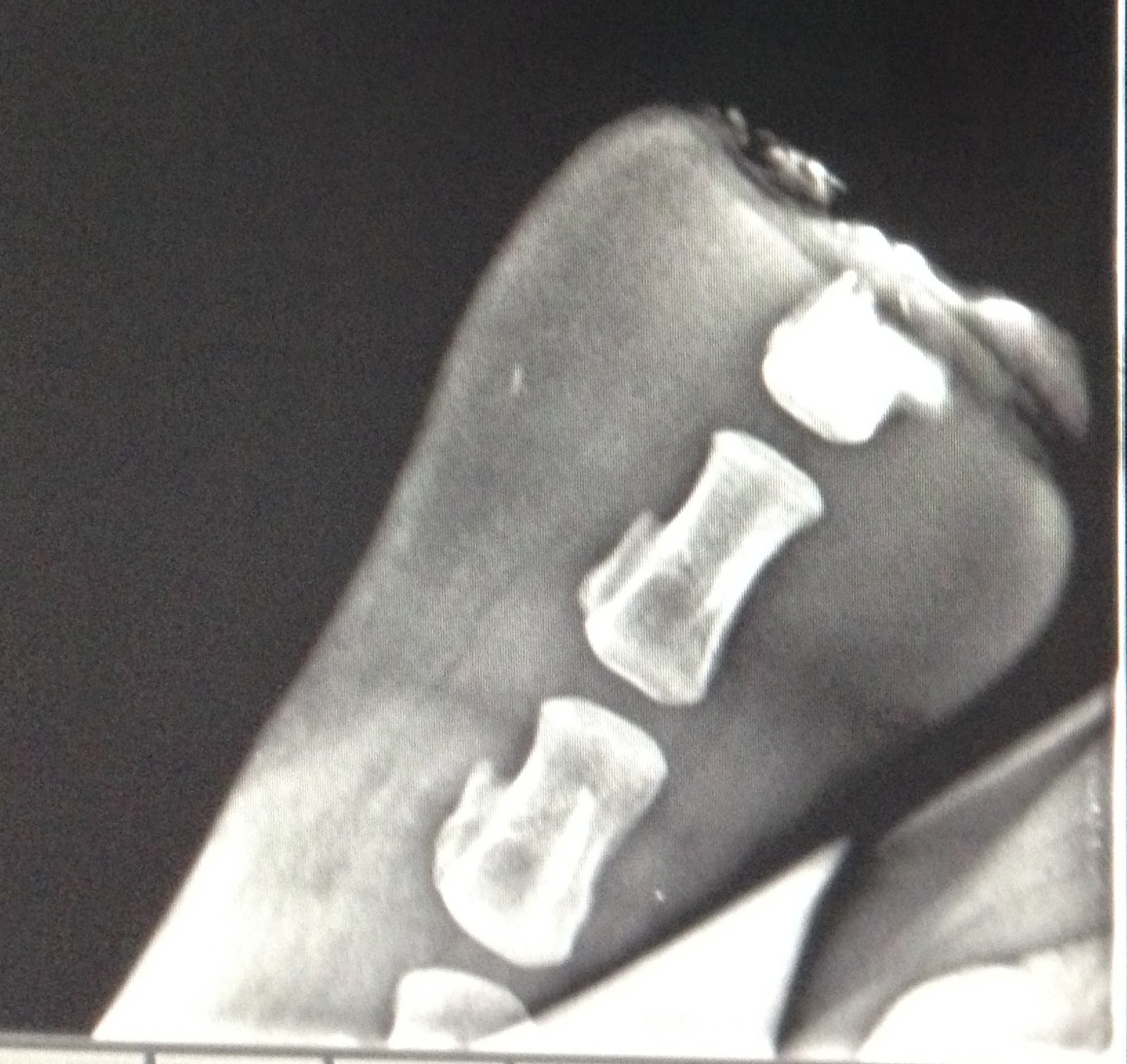
At the tip of the tail, there was part of a vertebrae remaining from the prior amputation attempts.

Dr. Hackett and Dr. Younkin removed the remainder of that vertebrae and a great deal of the granulation tissue in order to have enough skin to close over the end of the tail.


Closure with a Near-Far-Far-Near suture for tension relief, and then a simple interrupted pattern was placed in between the tension relieving sutures for apposition of the raw edges.

The patient will need to be on systemic antibiotics and anti-inflammatory drugs for several days to prevent further infection and minimize pain, but the prognosis for recovery is good!

The next couple weeks will bring several big changes to the clinic.  Dr. Younkin will be finishing his internship at BEH and heading off to Kansas State for a new position.  He has been a valuable source of information in my first month at BEH and we wish him only the best as he advances in his career.  A new intern who recently graduated from University of Pennsylvania will be starting next week, and BEH will also be welcoming a new board certified surgeon to the team from Tufts University, Dr. Patricia Provost.  I am looking forward to having even more people to learn from! 


Until next week!

Wednesday, May 28, 2014

Getting my feet wet at Clinton Veterinary Service: My first 2 weeks

I’m Valerie, a veterinary student from Michigan State University who recently finished second year and is looking forward to a summer of equine work. The first two weeks at Clinton Veterinary Service in St. Johns, MI flew by with remarkable speed. The practice currently has 6 doctors who largely work as a large animal ambulatory practice with some equine cases trailered in to the clinic occasionally. This summer I am primarily riding along with Dr. Cynthia Trombley, an equine vet who graduated from MSU in 1995 and co-owns the practice with her husband. From my first day onward, we’ve been quite busy. This time of year the majority of the calls are routine maintenance appointments: vaccinations, Coggins testing, health certificates for any horses traveling out of state, fecal floats and consultations on deworming regimens, and checking teeth for any needed dental work. In my short time here, the difference regular care and preventative medicine makes in the health of a horse is already readily apparent. With Michigan’s long winter, many people are just now transitioning their horses to pasture, which means we’ve had an increased number of colic calls. The majority of the cases have been gas colics, likely caused by the switch to fresh pasture, and have been successfully treated medically on farm. Fortunately few of the colic cases we’ve seen since I started needed surgical treatment, but our close proximity to MSU is helpful in the event surgery is warranted and the owners are willing to pursue that option.
                Dr. Trombley also sees a modest amount of reproductive work. Three days a week we visit a large Arabian farm to ultrasound mares. Ultrasound is useful for tracking a mare’s cycle, identifying follicles that are likely to ovulate, and confirming pregnancy. Unlike in cows, pregnancy can be diagnosed as early as 14 days by ultrasound. While I have yet to palpate or ultrasound a mare, I am becoming much more comfortable with interpreting what I see on ultrasound. The repro work is enjoyable and particularly rewarding when it results in a successful pregnancy. Since the decline of the racing industry in MI and the recession, there have been far fewer clients breeding horses these days, but we still have been out to several other farms to ultrasound mares, collect samples for uterine cultures, perform breeding soundness exams. The most gratifying calls are the new foal check up calls. I enjoy working with foals, but seeing the owner’s excitement over new life and new potential is very satisfying. Hearing owner’s talk with deep pride in their voice about the new foal that is the result of many generations of careful breeding and years of commitment to a breed or disciple is a good reminder that we as veterinarians may treat animals, but we do so in the service of people. We work to maintain the health of animals that bring many people a great deal of joy.

                I have seen and learned a great deal in the 2 short weeks here at Clinton Veterinary Service, and I’m looking forward to the weeks to come.


 A new foal visit to check out this nice little colt. It is also advisable to examine the placenta whenever possible after parturition.