Monday, August 25, 2014

The end of summer

As the summer comes to an end and school starts I thought I would write one last post to sum up the summer. The last horse show we attended was Youth Nationals in Albuquerque, New Mexico. It was a tough show for the horses because they travel a long way to get there and they are ridden a lot at the show. At this show we did our usual work to help the horses remain sound during the show, but we also had several medical cases to treat. These consisted of colics and pneumonias. The colics that we had were treated by giving I.V. fluids and banamine if the horse was far enough away from showing. There are certain drug rules for horses that are showing to ensure that no horse has an unfair advantage. We had to be careful of this when treating the horses. The horses that had pneumonia were given antibiotics, and sent to a nearby clinic. This was because they need more extensive care than we could give; they require the antibiotics at certain time intervals that we were unable to continue to do. Other than the medical cases, there were a few interesting lameness cases.

In one case the horse was a grade 4 out of 5 lame with a swollen leg. We brought the swelling down, and watched the horse move again. Once the swelling was gone the horse was the same grade of lameness. This caused us to perform other diagnostics. Dr. Ocull thought there was a potential tendon injury so she began with an ultrasound which showed a potential foreign body. We then took a few x-rays of the horse's fetlock to try and see the foreign body. The x-rays showed nothing, which made us believe the substance was not metallic. Luckily Dr. Wallis was with us and he is a board certified surgeon. He removed the foreign body with the horse standing. It turned out to be a wood chip, and the horse went on to compete in his classes.

After the horse show we drove back to Michigan and our work load slowed down. We did our normal routine calls, looking at lameness cases at different farms. We also had one more surgery day, where Dr. Hill worked up some referred lameness cases. Overall, I had a really enjoyable summer with performance equine. I learned a lot and got to see some really cool cases. It was a very hands on experience and Dr. Hill was a great teacher. It made for an interesting and fun summer.
Dr. Wallis removing the wood chip

The wood chip from the horse's fetlock. 

The wood chip on ultrasound. 

Ultrasound image of a normal lung. 

Ultrasound image of a lung from a horse with pneumonia. 

Tuesday, August 12, 2014

Wrapping it up

I can’t believe how quickly my time at Cleveland Equine has passed! During my final week, Dr. Robertson came in twice for surgeries. He performed two tie-back surgeries and one tie-forward. Tie-backs are used to treat laryngreal hemiplegia or “roaring”.  Roaring is a result of the paralysis of one or both of the arytenoid cartilages in the larynx (the left arytenoid is the most commonly affected.) The paralysis prevents the larynx from fully opening when the horse breathes, which can cause poor performance and a hoarse “roar” during exercise. The tie-back surgery uses a heavy suture anchored in the muscular process to hold the affected arytenoid open. Dr. Robertson also performs a unilateral ventriculocordectomy with the tie-back. This procedure removes the vocal cord and ventricle on the affected side to create more room within the airway.
            The tie-forward procedure is performed as a treatment for dorsal displacement of the soft palate. As I have written about a couple times, there are many treatments for this condition such as lasering the soft palate or performing a myectomy. Tie-forwards are generally more successful, but more invasive and expensive because it requires general anesthesia. In this procedure a strong suture is strung through the basihyoid bone and the larynx, then tightened to bring the two structures closer together. By elevating and bringing the larynx forward, the epiglottis creates a tighter seal with the soft palate, which prevents future displacement.
            Both Dr. Paradine and Dr. Berthold took time out of their busy schedules this last week to give me some practice with joint injections. Dr. Paradine pulled out the old stud kept at the clinic for teaching purposes and guided my through injecting the distal intertarsal and tarsometatarsal joints of the hock as well as the fetlock joints. On my first tries, I gained a better understanding of what “incorrect” felt like, but I was able to successfully inject at least one of each joint. All that practice paid off when Dr. Berthold had me inject the same two lower joints on his daughter’s riding horse; I was able to do so with very little trouble. He then showed me how to inject the stifle joint and had me give it a try.
            I can’t believe how incredibly supportive and welcoming the doctors and entire staff have been here at Cleveland Equine. They truly made my summer experience remarkable; I can’t believe how much I learned in such a short period of time. Whether it was on the road or in the clinic, each vet I worked with took the time to answer my questions and offer helpful pieces of advice. I cannot think of any better way to spend a summer.

Friday, August 8, 2014

Vesicular Stomatitis and Drug Induced Autoimmune Hemolytic Anemia

Hey all,


These past few weeks have certainly been eventful.  Colorado has had several cases of vesicular stomatitis (VS)- a reportable viral disease that causes fever along with vesicles and ulcerated lesions along the gums, tongue and coronet bands of horses.  VS is a mild disease in horses, but preventing the spread to other species is a great concern.  VS can be transmitted to cattle, swine, sheep and other species where the signs are indistinguishable from foot-and-mouth disease without testing.  Severe economic losses can result as oral lesions prevent livestock from eating, and lesions on teats lead to decreased milk production.  In addition, VS is occasionally transmitted to humans where it causes flu-like symptoms.
VS is spread by insects and contact with vesicular fluid (through direct contact, shared drinking water, etc).  A barn with VS is placed under quarantine, and the affected animals are isolated with separate equipment and water.  Even the clinic would be quarantined if a horse with VS were to be unloaded on the property.  To prevent that from happening, the doctors initiated a policy whereby no horse is allowed off their trailer until it has been inspected for VS and shown no signs of the disease.  The veterinarian doing the exam wears coveralls and exam gloves to prevent themselves from being contaminated.  Here is the link to the APHIS website about VS if you want to know more:  http://www.aphis.usda.gov/wps/portal/aphis/ourfocus/animalhealth/sa_animal_disease_information/sa_equine_health/sa_vesicular_stomatitis/ct_vesicular_stomatitis/!ut/p/a0/04_Sj9CPykssy0xPLMnMz0vMAfGjzOK9_D2MDJ0MjDzdgy1dDTz9wtx8LXzMjf09TPQLsh0VAZdihIg!/


We recently had one of the most interesting cases of the summer.  An older gelding came in for colic and went to surgery to correct a colon torsion.  The horse began to recover well in ICU, and it seemed he was going to be just fine  A few days into it, though, he developed a mild fever.  The veterinarian in charge suspected an infection and placed him on the cephalosporin antibiotic Naxcel.  Not long after that, the his condition began to deteriorate and his urine became reddish in color.  He developed a rare reaction to Naxcel in which his own immune system began lysing his red blood cells.  He was switched to the fluoroquinolone antibiotic enrofloxacin and placed on corticosteroids to depress his immune system and prevent further destruction of his erythrocytes.  Before beginning the enrofloxacin, an abdominocentisis was performed and fluid collected for cytology and culture.
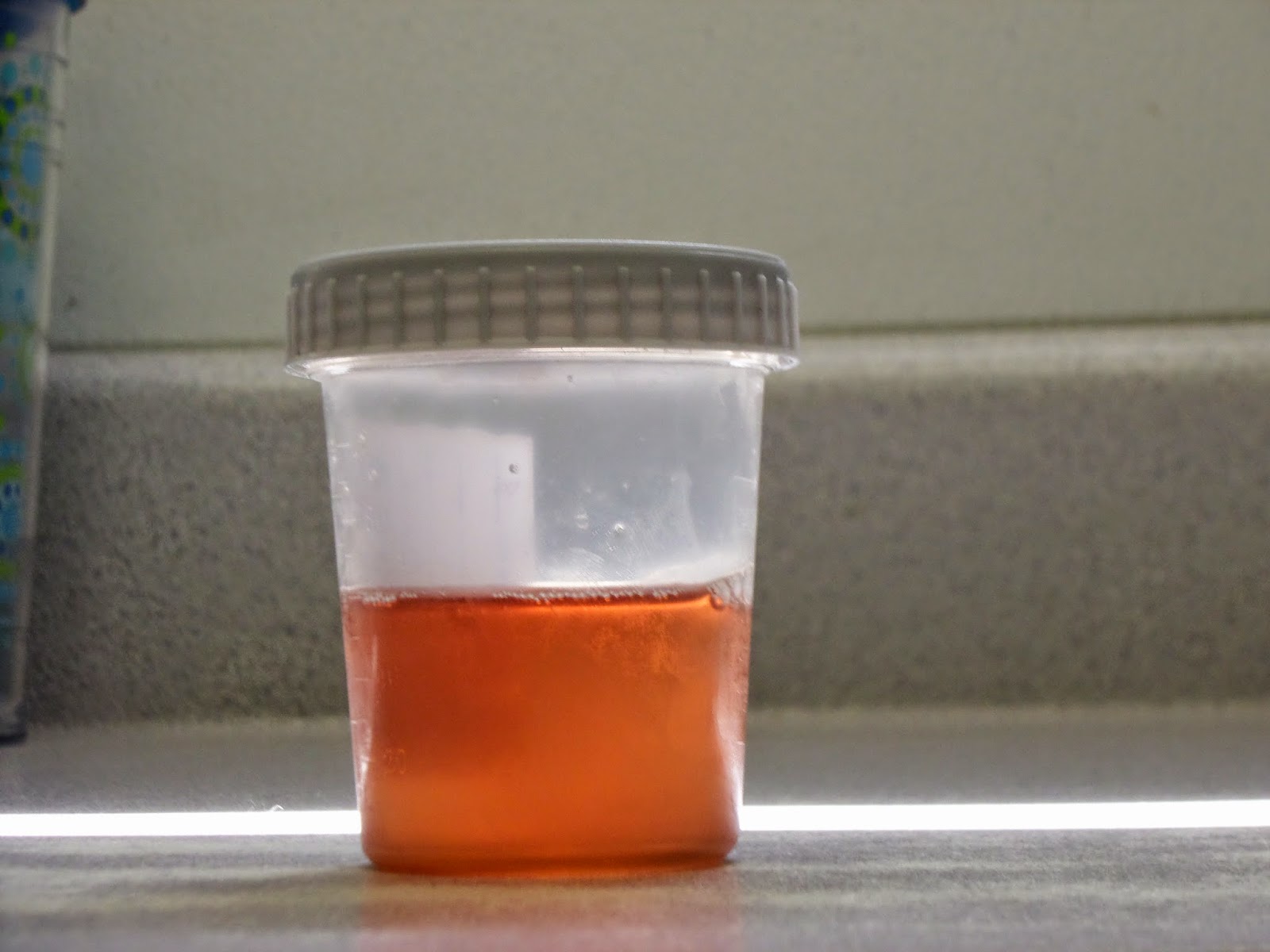
Here are some images of the horse's urine and abdominal fluid from a day or two after stopping Naxcel:

The reddish color of the urine is caused by the presence of hemoglobin from the broken down erythrocytes.


The abdominal fluid was very dark red like old blood.  I did not learn the results of the cytology and cell culture, but the veterinarian suspected that this was blood left over from surgery. 


My twelve week tour at Littleton Equine Medical center is at an end.  I have truly loved being here.  I had limited experience with horses in the hospital setting before coming here, so working in the ICU doing treatments and taking parameters was a great benefit to me.  I also learned a lot from the doctors about colic work up, lameness diagnosis, and how a clinic should respond to contagious disease outbreaks.  I have a lot more to learn, but I feel much more confident and excited about going into clinics this winter.

Monday, August 4, 2014

Weeks 10 and 11 at SVEC


            With Dr. Williams now working at Saginaw Valley Equine Clinic, there have been no shortages of surgeries going on.  A 16 year old stallion presented with one testicle about 3 times the size of the other.  The owner still wanted to be able to use him for breeding, so she elected to only have the 1 affected testicle removed.  At surgery, the vaginal tunic was already open suggesting trauma, although, there was no history of trauma that the owner was aware of.  Once the testicle was exteriorized, it was clear that more was going on.  The spermatic cord was very edematous and there were 3 small, hard masses on the testicle surrounded by scar tissue.  The testicle was sent to pathologists for examination and I am curious to see what the results come back as.  The clinic now has an arthroscope/ laproscope and we had our first ovariectomy this week.  The mare is 2 years old and exhibits dangerous behavior towards people.  Assuming her behavioral issues were hormonal in nature, this surgery will hopefully help resolve those problems, though it can take about a month post surgery to see a change. 
            One of our medicine cases from about six weeks ago came back in and unfortunately did not have a good outcome.  This 4 year old gelding was sick as a foal and has never quite been a normal horse according to his owner.  He chronically colicked at least every 3 months, was thin, and would get edema in the throatlatch region due to low protein.  When we scoped him in June, he had horrible stomach ulcers – there were large, deep ulcers all over his stomach but especially near the margo plicatus and hyperkeratosis in between the ulcers.  He went on a course of sucralfate and gastrogard and when we checked him near the end of his month long treatment his stomach looked great, with all ulcers completely or very closed to being healed and he had gained quite a bit of weight.  However, when we checked him after 2 weeks off of treatments, his ulcers had already returned and he had another colic episode.  Unfortunately due to the severity and chronicity of his issues at such a young age combined with the owners financial situation, the decision was made to euthanize the horse.  It was sad to see a young horse go but I definitely think it was right decision for this horse and this owner. 

Saturday, August 2, 2014

The last few weeks at RREH

    Howdy! Just wanted to take a moment and say that this has been such an AMAZING and REWARDING experience. I have learned so much and seen so many things during my time here, as well as have established relationships here that will last a lifetime. I am so thankful for this opportunity and cannot wait to do it again next year! 
    Anyways, onto the last few exciting cases I witnessed! To start things off, we had a teaser stallion come in due to a couple fractured incisors. The doctors came to the conclusion that he may have caught himself onto the fence, or some other trauma that did not involve much soft tissue around it. 


As you can see, Dr. Embertson wired the teeth together to that they can heal in their proper place

     For our second interesting case, we had a yearling come in with a giant laceration on its knee after a very loud and crazy thunderstorm. Dr. Embertson did a great job stitching this guy up!


   Last but DEFINITELY not least, is by the far the most interesting procedure I have witnessed done in surgery this summer. Dr. Bramlage performed an arthrodesis on a stallion last week. Equine arthrodesis is a medical procedure in which fusion of equine joints is performed through surgical, chemical or ethyl alcohol methods. Surgical arthrodesis involves destroying the articular cartilage surrounding the joint using a laser or surgical drill, and applying a bone plate or placing a bone graft between the joints. The joints are aligned into a stable, weight-bearing position and screws are placed in various positions depending upon the individual joint. Surgical arthrodesis results in joint fusion by removing the cartilage and allowing the bones to fuse together. 
    WARNING: these next images are very graphic and not for a weak stomach!


The leg is then casted and the horse stays at the hospital for approximately 30 days under close watch.


Thanks again to everyone who took the time to read my blog!