Tuesday, June 30, 2015

Week 6/7 Surgery RREH

Hello from Kentucky and it has been extremely busy these last 2 weeks! An exciting surgery that was performed was an arthrodesis by Dr. Bramlage. An arthrodesis is a surgical immobilization of a joint by fusion of the adjacent bones, resulting in bony ankylosis. This arthrodesis was performed on the left front fetlock due to a racing injury that occurred to the proximal sesamoids and the areas between the third metacarpal bone and the proximal phalanx, also known as the cannon bone and long pastern bone. Dr. Bramlage created his own condylar fracture to enter the joint space. By creating his own fracture he was able to reduce the stress and damage on the surrounding soft tissue. After entering the joint space the articular cartilage was removed to facilitate bone growth and remodeling to fuse the joint. The fracture he created was then fixed with screws and then 2 plates were placed to stabilize the joint. When the surgery was complete, the horse was placed in a cast with a drain tube and will remain in the hospital.
Warning Graphic Pictures: 

The initial incision is made and it is time to create the condylar fracture to enter the joint.

The joint space is entered and it is ready to have the articular cartilage removed and subchondral bone exposed. You can see the third metacarpal bone to the left of the picture.


Removing the cartilage with a bone saw. He used this earlier to make the condylar fracture.


The first plate is in place and screws were placed to hold it.


 The last picture is of Dr. Bramlage manually hand driving in the screws after using a drill to make sure they are securely in place.

 It was such an amazing experience to watch Dr. Bramlage perform this surgery! Until next time, thanks for reading!
Jackie Tobias

Sunday, June 28, 2015

Lymphangitis, Pericarditis, and Colics at Littleton

The last few weeks at Littleton Equine has been bust with many interesting cases. These include colics, lymphangitis, and pericarditis.

Lymphangitis is a severe form of cellulitis and is the swelling and inflammation of lymphatic vessels. The damage to these vessels is usually caused by a bacterial infection. This condition usually occurs in a rear leg. The horse will present with an extremely swollen leg, sometimes 2-3 times its normal size, a fever, pitting edema, lameness, a wound, and skin damage. Many of these horses have a loss of appetite and be very painful. These horses need to have multiple cold hosings a day to help reduce the inflammation and swelling.  This also helps wash off oozing serum from the skin so it does not irritate the skin. We were able to use a Game Ready system to apply pressure as well as cold therapy to the leg. She was also hand walked multiple times a day to help move the swelling out of the leg. Pain medications, such as anti-inflammatories, and antibiotics were administered to keep her comfortable and help treat infection. Having lymphangitis once, a horses is more likely to develop it again in the same limb due to impaired immunity. There may also be residual swelling after called lymphoedema.


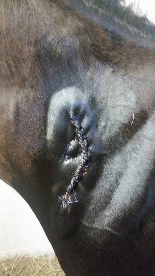
Dr. Christakos went to a farm to do a health certificate. Upon her exam, she found the horse to have an extremely elevated temperature and muffled heart sounds. When he arrived at the hospital an ultrasound was performed by Dr. Toppin. The paint gelding was found to have a large amount of fluid around his heart known as pericarditis. Pericarditis is pretty rare in horses and can be caused by a septic infection or it is idiopathic. Dr. Toppin drained the fluid from the pericardium and lavaged the pericardial sac with saline. He was placed on stall rest in ICU to monitor him. His heart rate remained elevated during his stay and occasionally had AV block. His appetite was great during his stay and he enjoyed dumping his water bucket all over his stall during his boredom. 
Location of pericardiocentesis 

We have had multiple colics in the past few weeks. One of the colics was a donkey gelding. He presented for being lethargic, dribbling urine and decreased appetite. He was a very stoic boy who did not show his pain. He was admitted to the ICU for fluids, monitoring, and refluxing his nasogastric tube. His heart rate slowing elevated as high as 140. The owner elected to take his to surgery. In surgery, Dr. Murray found him to have a gastric impaction, a severe small colon impaction, and a dorsal displacement. The small colon was massaged to help move the feces along and the large colon was put back in place. 

I went out with Dr. Toll to field calls. We went to a colic of a Quarter horse gelding. The previous day he laid down on the owner during a trail ride. Dr. Toll was wondering about the possibility that the gelding had tied up but he walked extremely well when we arrived on the farm. He was not interested in food. She passed a nasogastric tube, but we did not get any net reflux. She however felt that the tube did not fell right and was worried about gastric impaction. Upon rectal examination, dry feces was found in his small colon. She recommended the owner to transport him to the hospital for further work up and monitoring. Before transporting him to the hospital, we gave him oral fluids with electrolytes through his tube to help with his dehydration as well as break up the potential gastric impaction. His blood work at the hospital showed elevated liver enzymes and a elevated PCV. After a few days of fluids, he had still not passed any feces and his liver enzymes remained high. Liver disease was a concern.  He remained very stoic in ICU as well and his heart rate remained within normal limits. He was taken to surgery to explore his abdomen. Dr. Murray found him to have a dorsal displacement. Following surgery he did very well and was able to return home after about a week and a half in the hospital. 

Another colic did not end so well. After surgery, the 27 year old TB gelding continued to reflux fluid due to enteritis. He had a adverse reaction to a medication and he became a danger to be around. We were unable to control his adverse reaction and he unfortunately had to be euthanized. I had the opportunity to help Dr. Rice with the necropsy. The poor old man had displaced again and his small intestines were extremely angry. 
Lipoma in the abdomen

Proximal Enteritis Case

We were called out to a farm yesterday to see a 10 year old Standardbred gelding “Andy” who had a fever of 103 deg. F, dark urine, and depression. When we got there, he was a little sweaty and looked like he had been rolling. On physical exam, his heart rate and respiratory rate were elevated (52 beats/min and 36 breaths/min). He was approximately 5-7% dehydrated. On rectal palpation, he had scant manure and mucus, and his cecum was moderately distended. When we passed a nasogastric tube, we obtained over a gallon of yellow, foul smelling bile from his stomach. With banamine, sedation, and several liters of IV fluids, the gelding’s heart rate, respiratory rate, and temperature decreased. His attitude also improved and he drank some water. However, he continued to spontaneously reflux out of the nasogastric tube intermittently.

The veterinarian presumptively diagnosed proximal enteritis (aka anterior enteritis or duodenitis -proximal jejunitis). This is a type of colic caused by a functional obstruction. Inflammation of the duodenum and proximal part of the jejunum lead to ileus – cessation of small intestinal motility. The stomach and small intestine hypersecrete fluid which has nowhere to go and can eventually rupture the stomach if a nasogastric tube is not placed. The horse can become hypovolemic due to loss of fluid into the GI tract. The cause is thought to be bacterial, either Clostridium difficile or C. perfringens or Salmonella spp, but is not well understood. There is a significant risk of endotoxemia or endotoxic shock occurring due to leakage of bacteria from the gut.

In this case, we suspect the proximal enteritis was initiated by a change in diet. The owners were running low on hay so they switched to a diet of primarily grain and a few hours turnout on pasture. After initial treatment, we referred this horse to a specialty hospital for in-patient care. He will need large amounts of IV fluids for the next few days, gastric decompression through the NG tube every two hours, IV antibiotics and pain medications, and his feet iced to prevent laminitis. We anticipate that “Andy” will have a good prognosis, since despite the severity of his condition it seems that it was caught early and the owners were able to take him promptly to a referral facility. His owners, an Amish couple, use him for driving and he is their only horse, so we wish him a full and speedy recovery!

end of week 6 at Browns

Rain keeps pouring down but we keep working!
Do you guys know the solution to fixing a "roar" in a horse, or otherwise known as laryngeal paralysis? I bet the first thing that comes to mind is a tie back in which suture is placed to physically "tie back" the paralyzed arytenoid. Tie backs are performed on horses that undergo strenuous exercise and need maximum air intake at all times, such as thoroughbred racehorses. However, here at Brown we see a lot more pleasure horses as well as draft pulling horses. Since these horses don't need maximum oxygen intake at high speeds, Dr. Brown performs a ventricular chordectomy on all of them. This procedure isn't as good at opening the airway back up but it gets rid of the roar nonetheless, and for horses that aren't running high speeds its the perfect choice. It is a standing procedure with no suture placed, which makes it safer on the horse as well. Using a scope to visualize what is being done, Dr. Brown makes an incision between the jaw bones, in the throat, right at the level of the arytenoids. He then takes out the saccules that line the ventricles, as well as removes the vocal folds; when this tissue heals, it scars down, creating tension on the paralyzed arytenoid, to keep it from flapping in the airway and causing noise. He removes both sides to assure a tight scar throughout. 
In this image, you can see what is being done on the screen; there is a hemostat clamping off a saccule, ready to be removed! We do this surgery about 2-3 times a week, because he is very efficient at it and the success rate is tremendous! 

Another fun case was a horse that presented 3 legged lame due to a quarter crack. This crack was very severe and his lameness was well justified! Dr. Brown cut away the dead hoof and passed wire from side to side to try and bring the crack back together. He also filled the back side of the heel with epoxy glue (which hardens into plastic-like consistency) to make sure that the horse is stepping on his heel more to prevent rotation of P3 while he is healing. 


Thanks for reading this week!

Monday, June 22, 2015

Update from Texas

The horse show in Minnesota went well. It was a little slow again, but we didn't have any sick horses which was nice. The weather up there was also a nice change from the Texas heat! The last couple days of the show we did lameness exams and treated a few horses for a couple barns. On our way back down to Texas we stopped in Missouri to work on some horses at two farms. At one of the farms we saw a horse who had had upward fixation of the patella and had to be treated by cutting the medial patellar ligament. We took radiographs of his stifles to make sure he wasn't getting changes in the joint from the procedure and they looked pretty good; the only abnormality was that the patella sat a bit more lateral on the caudal cranial view. At that farm we also treated one horse's navicular bursas because of known changes to the navicular bone. This injection is pretty cool; it is a blind stick and then a radiograph is taken to confirm the location of the needle. I got to shoot the radiographs for the procedure. From there we went to the next farm which was more lameness exams and treatments. We finally made it back down to Texas for the rest of the week. At one of the farms we did a pre-purchase. We also pulled blood to get PRP for the treatment of a superficial digital flexor injury. The tendon was treated ultrasound guided and done at the farm because Dr. Wallis has a PRP machine on the truck. At the next farm we went to we saw a horse who had lacerated his upper eyelid. It had already gotten stitches, but we were asked to look at it. We cleaned it up and Dr. Wallis felt it could use one more stitch so he added one near the medial canthus (the corner of the eye). That ended our week, and now we are headed back to Missouri to look at horses and get them ready for Region 11 next week. That's all for now.

Jenna

Saturday, June 20, 2015

Week 6 at Rood and Riddle

I can not believe I am already halfway done with my time here at Rood and Riddle! I can not even keep track of all the amazing things I have learned and done since I got here. But I have been trying to get as many pictures as possible to show you all the things I have been able to see and do.

 These first two pictures are from a Saddlebred colt who ripped his shoe off and took a large portion of hoof with it. I thought this was really cool because on the inside of the shoe, the hoof wall was still attached and you could see the lamellae still attached.


This is from a young foal that ran a fence board and had about a 4 inch wide gash in his neck. Dr. Friend did an awesome job sewing it back together.


In these pictures I am sewing a caslyx on a Thoroughbred mare who has just been bred. These are not as common as they used to be, but some farms still caslyx every mare after they have been to the shed. So far I have done about 5. I also found my first pregnancy in a mare who was 14 days from ovulation! It is amazing how exciting it is to see the little black circle on the screen. My ultrasound skills are still a little rough but I am getting better everyday.


This big guy is from a pre-purchase we were doing where Dr. Friend needed to make a phone call so the owner graciously let Kelsey and I have some fun with their horse! Everyone needs to have some fun every once in awhile!


 This picture is from a barn that got rotovirus where we had to quarantine the whole barn to make sure the new foals would not get infected. The plastic coveralls are unbelievably hot in this Kentucky heat and humidity. 


Castration day! One of our regular farms decided to castrate 40 of their stallions and we have been doing them in sets of 10. This day was the first set and I was able to help with one and then do the last 3. 
Last week I went to visit Jackie in surgery for a day and got to hangout and see a few arthroscopies and fracture repairs. It is always fun to go back to the hospital and see how much of a difference there is between field and hospital medicine.
                              

The last of my adventures end with a party at Churchill Downs that Nicole and I were able to attend. It was a meet and greet with American Pharoah and Victor Espinoza and their trophies. It was wonderful to get to hangout with such an amazing athlete!!!

That is all for now! Happy Father's Day!!

Friday, June 19, 2015

week 5 at Brown Equine

Busy busy all day is the name of the game!
Typical appointments come and go, keeping us on our toes at all times.
I wanted to share an awesome, and very DISGUSTING case we had come in today. This horse presented to us for a facial swelling on the right side of his face.
As you can tell, this is quite a large, and very easily seen mass. Before even doing anything, Dr. Brown was more or less certain of the diagnosis. It was a paranasal sinus cyst. Why so certain? because no other commonly seen nasal sinus lesion would cause a facial deformity as extreme as this one. After doing a standing nasal flap surgery, in which we drill a hole in the front of the head at the area of the frontal and maxillary sinus, we emptied all the cyst contents and packed it with betadine pads which will be removed in a few days. Dr. Brown hopes that his face will return to a more or less normal shape with time. I have attached a video for all to see of us opening up the sinus and getting rid of the cyst itself but be warned!!!! This video is not for the weak stomach, because is PUS PUS PUS! For all those who like that sort of thing, ENJOY!


Thursday, June 18, 2015

Week four and five at SVEC

Sorry it has been a while since my last post!  Time here at Saginaw  Valley Equine Clinic is flying by.  I can't believe we are in the sixth week of our fellowship here.  Over the past couple of weeks, I have had the opportunity to work on my handling skills with plently of fractious patients.  We currently have a lot of post-surgery patients in the clinic, giving me opportunities to work on my treatment administration.  While I have not had many opportunities to scrub in on surgeries, I did get the chance to scrub in on an arthroscopy on a horse with a steroidal joint infection following a joint injection.  I absolutely loved being about to view the joint from a different angle than the usual radiographic views.  Although the past few weeks have been trying at times, I am more sure every day that I want to work in equine medicine for the forseeable future and can easily see myself specializing in sports medicine.

Hopefully this week I will write my blog post in a more timely fashion so I can give more case details!

~ Elaine

Tuesday, June 16, 2015

Week 5

Hello Everyone from RREH!


 This past week was a crazy week in surgery with a variety of colic surgeries including a small intestinal volvulus, large colon volvulus, and an impaction all going to surgery. A really interesting case was a foal with an umbilical abscess. The infection had traveled in the umbilical vein which became enlarged and thickened due to the prominent infection. Above are pictures of the surgical removal of the abscess and the infected vein. The last pictures are of Dr. Woodie untwisting the large colon volvulus during the colic surgery! I was also lucky enough to ride with Dr. Brown and Dr. Friend to learn what it is like to be an ambulatory veterinarian in beautiful Kentucky! With Dr. Brown we spent the majority of the day doing pregnancy ultrasounds and checking on yearlings with various lacerations and hematomas. We also scoped a yearling to evaluate his airway because he was having trouble breathing in the pool post training sessions. With Dr. Friend we evaluated different lameness cases and long term management of these horses. Everyone as been so incredible about teaching and I can't believe how much I have learned!
Until next week!!
Jackie

EAS: ultrasounding hearts and seeing AP!

After working religiously on my research this past week, I finally got to put my hands on a real live racehorse and work on my ultrasound skills! With the help of Dr. Bo Rainbow, I was able to get some good images of the heart.


In other events, Kentucky has proven quite exciting lately, with the return of American Pharoah to Churchill Downs following his Triple Crown victory. Horsemen and racing fans alike have been enamored with the impressive colt and he did not disappoint when the crowds of admirers (myself included) flocked to Churchill Downs this past Saturday night to see him parade around the paddock. He truly is a special horse and I am so excited to be working in the Thoroughbred industry during his reign. Hail the Tail!!!


Best,
Nicole