Week ten started out with 13 lameness exams in one day! Twelve
of those lameness exams were for one of the standard bred clients that we work
with. Usually on Mondays or Tuesdays we work on their race horses and conduct lameness
exams and identify the issue and treat them accordingly. The client wanted
their horses to be ready for the Delvin Miller Adios Pace for the Orchids at
the Meadows. The Adios is a horse race for three-year-old Standardbred colts
and geldings that occurs annually at the Meadows Racetrack in Washington
County, Pennsylvania. This is a final for the horses and it is a one-mile long
race. During the lameness exams we can identify different issues the horse is
having by having them trot in circles to the right or left and also flexing the
horses limbs. You can do upper and lower limb flexion as a way to identify what
is the source of lameness in a horse as well as using hoof testers. For
example, when the horse is positive (has a change in the degree of lameness) to
both upper and lower limb flexion in the hind limb, the suspensory ligament can
be the issue. You can then clip the area and use an ultrasound to examine the
suspensory ligament and look for any tears or abnormalities within it.
Sometimes nerve blocks are utilized when the site or source of the pain is unclear.
If you are able to block the horse out successfully and the horse is then sound
upon re-examination, you have localized the pain and can therefore use other
diagnostic procedures more effectively both medically and economically to
identify the cause of the lameness and treat it appropriately. Being able to
work with Standardbreds was not something I had envisioned for myself as a future
career path and I am glad I had the opportunity to do so. This gave me the chance
to experience a different career for myself and working with clients like this allows
me to see that it is a definite possibility.
Later on in the week, there was a patient that came in due
to a fracture at its first phalanx (P1). The horse was competing at a barrel
racing contest when it fractured its P1. We took some x-rays of the patient in
order to visualize the fracture and see if it could be repaired surgically.
Here are some pictures of the x-rays we took:


The owner wanted to move forward with surgery, so we placed
a catheter, administered the pre-anesthetics and took the horse to surgery. We
had an x-ray machine in the operating room in order to take images of the
procedure and confirm placement of the screws and that adequate compression was
generated. The surgeon needed an extra hand and I was fortunate enough to scrub
in for the surgery and hold the horse’s limb in position while he worked. I
held the limb in a curled position, similar to curling a weight, for an
extended period of time (sorry I didn't get any pictures of this, I had my hands full!). It was safe to say it was arm day for me, no need to
go to the gym! We placed all of the screws, put a cast on the horse and put the
patient in the recovery stall. The horse recovered wonderfully from anesthesia and
stood up nicely. The horse did not arrive to the clinic until 5 p.m. that evening
so surgery did not start until 8 p.m. The surgery lasted about 5-6 hours and recovering
horses takes time. We did not leave the clinic until 4 a.m.! While it sounds
tiring, it was worth it! I can honestly say there really isn’t anything like
surgery. The horse is still at the hospital and recovering. The cast will not
come off for another couple of weeks, which the horse is less than enthusiastic
about. Before I left the horse started trying to remove parts of their cast,
but to get them to stop doing that, we placed a half limb bandage above it and
put paste on the outside of the bandage to deter them from eating the bandage.
This proved to be effective and the horse is no longer biting at their cast.
Here are some of the pictures taken during surgery and after we placed the cast and screws:


We also had a couple of horses come in for ventral
cordectomies (VC). These horses were experiencing exercise intolerance and we
scoped the horses to see if they were paralyzed or not. The horses were both
experiencing left recurrent laryngeal paralysis and needed to undergo the VC
procedure. The area was clipped, scrubbed and blocked. We utilized the scope
and placed it through the patients nasal passage in order to adequately
visualize the area while the surgeon removed the saccules and vocal cords of
the horse. The patients recovered well and were sent home the next day.
Then we had a horse come that was three legged lame. This means that the horse was only functional on three legs. It would hop instead of putting weight on its front right leg. We took the bandage off of the horses hoof and found that the horse had an abscess that burst through the toe of the hoof and was infected. This was causing the horse a significant amount of pain. In order to debride the area the surgeon decided to use medical maggots. Medical maggots are disinfected maggots used to debride wounds and clear infected tissue without harming healthy tissue. The maggots are applied to the area and bandaged so that the maggots stay in the wound and do not migrate away from the affected area. After about 3-4 days the wound is re-checked and additional maggots are placed if needed. In addition to the maggots we consulted with the Ferrier in order to see what we could do to make the horse more comfortable. When we walked the horse, it would put weight through the front right hoof when going up and down hill. When she would go back towards the barn, she would begin to limp again. The slope of the hill was causing her to be uncomfortable on the lateral portion of her hoof. This meant that she would be more comfortable with a wedged shoe that was placed on the lateral aspect of her hoof. To help the Ferrier trim their feet we took x-rays to show him how far he could trim the horses feet. We also ordered easy
riders to help her be more comfortable and provide support to the other hoof. The horse stayed at the hospital until the maggots were gone and was discharged home.
Another horse came for a lameness exam and before the exam even begun the horse's hind legs looked like it was dragging and it would sometimes even kick out. (For my fellow first years, it looked almost exactly like the video Dr. Manfredi showed in the Stifle lectures). This horse had a locking patella. In order to fix this problem the doctor decided to fix it surgically by doing a medial patella ligament splitting on both legs. What the procedure involves is cleaning the area, anesthetizing the horse as well as applying local anesthetics to the area. Once this is done the surgeon takes a blade and makes about three punctures with a scalpel blade in order to make the medial patella ligament thickened and enlarged. This helps to make it less likely that the medial patella ligament will get suck over the stifle and allows the ligament to be more fixed in space. This will help make movement for the horse easier and will decrease the instances of the horse dragging its toe and kicking out.
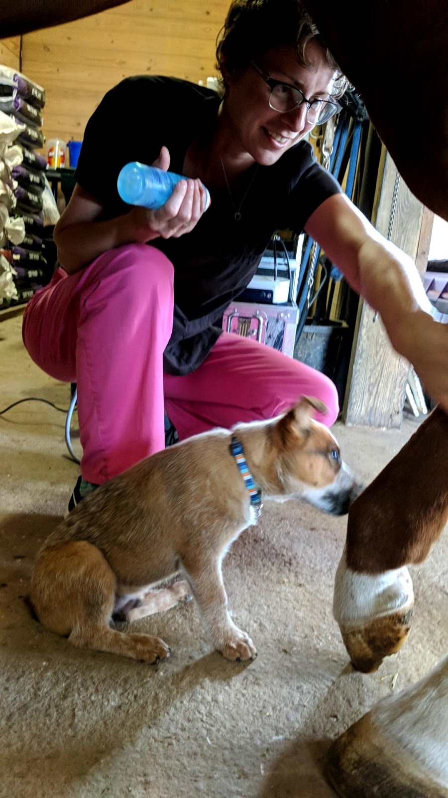
I was able to go out to one of the neighboring farms with the vet techs to shock wave a couple of horses. I was able to bring the machine with us in order to shock the horses suspensory ligaments and hocks. This was done on some racing horses in order to help stimulate healing in affected areas. I even had a helper with me (pictured below)! Normally you would do a lameness exam first before doing shock wave therapy, but these horses had already had lameness exams before and were in the middle of their treatment cycle.

Over these past four weeks I have been able to gain more experience placing IV catheters, administering IV medications, taking x-rays, working up colics, performing PD and abaxial nerve blocks, palpating for injecting joints, prepping and recovering horses from surgery. In between the appointments and surgeries I have been able to help with inpatient care and gain more experience with charting for patients as well as learning how to calculate dosages for patients.
I cannot believe that the 12 weeks for this fellowship is already over. It is a bitter sweet ending to be honest. I have had long days and many sleep deprived nights, but I would not change it because it has taught me many things about the profession and myself. It has allowed me to get a small glimpse at what an internship is like and if I am up for the task. It has shown me what vet med is really like and what kind of veterinarian I want to become.
I had two goals when I started this job: to work hard and learn as much as I could about equine medicine. I believe that I accomplished said goals, granted I still have more to learn about equine medicine, but I believe that I am off to a good start. I cannot thank the staff at Brown Equine enough for everything they have taught me and done for me this summer. I am forever grateful for all of the hands on learning opportunities that I was afforded here. Because of them I feel more prepared for clinics and I am more confident in my abilities. Thank you again for all of your help, I will miss you guys!
Until next time!
Shelbe