Wednesday, June 27, 2018

Clinton Vet Service: Week 4

Another week filled with more interesting cases has come and gone at Clinton Vet.  I would have to write a small novel in order to encompass all of the unique cases that I have had the chance to be a part of during my time with Clinton.  I have chosen two of my favorites to review in this weeks post.  Thank you all for following along this month, enjoy!

Eye ulcers can be a frustrating and drawn out battle for horse owners.  They can have multiple causes, including abrasions, foreign body, and neoplasia.  This particular gelding has been battling ulcers on and off for almost two years.  In the past he has gotten them in the winter time, this being the first time he got one in the summer.  A week prior to our first encounter, one of the associate veterinarians had performed a corneal scraping and sent it into MSU for evaluation.  Surprisingly the sample came back positive for squamous cell carcinoma, with a note that a full thickness biopsy would be needed to confirm.  The owner was extremely worried that a positive cancer diagnosis would result in the loss of his eye.  After looking at the eye we did not suspect that it was squamous cell, as there was no appearance of outer eye involvement or cancer-like behavior. 


Ulcer taking up stain
on our first visit


No stain uptake after
biopsy and treatment
        We began our exam by looking at the eye with an opthalamascope and light.  Then we stained the eye fluorescein, a dye that is taken up by damaged or inflamed cells in the eye. So we put him back on daily ulcer treatment, in this case a triple antibiotic, atropine, and an anti-fungal just in case.  The owner was willing to bring him into MSU for an ophthalmology consultation and biopsy so we got him in as fast as possible.  MSU took a biopsy and instructed her to continue treatments.  All of the tests came back negative; which was both encouraging and frustrating.  We were back the drawing board as there was not evidence of bacteria, fungus, a virus, or cancer.  Two weeks later we returned to the farm to a welcome sight.  The eye no longer took up stain and the biopsied portion was healing well.  For now we are discontinuing treatment and crossing our fingers that we have cleared his ulcer woes once and for all.

Cytology showing abnormal lymphocytes
The next mare that we visited was on the schedule for "bumps on her body" and weight loss.  A long time client of Dr. Cynthia's, the owner just wanted to trouble shoot what could be causing her problems.  The mare is older and has been struggling with weight loss for some time.  Dr. Cynthia palpated the area, near the mares right flank, and noted that it was well attached to the underlying tissue, a sign that it might be neoplastic in origin.  We took a fine needle aspirate of the area and brought it back to the clinic for cytology.  Unfortunately, we noted a number of abnormal lymphocytes.  Our most likely diagnosis was cutaneous lymphoma.


Taking a punch biopsy
The "lumps", more on underbelly
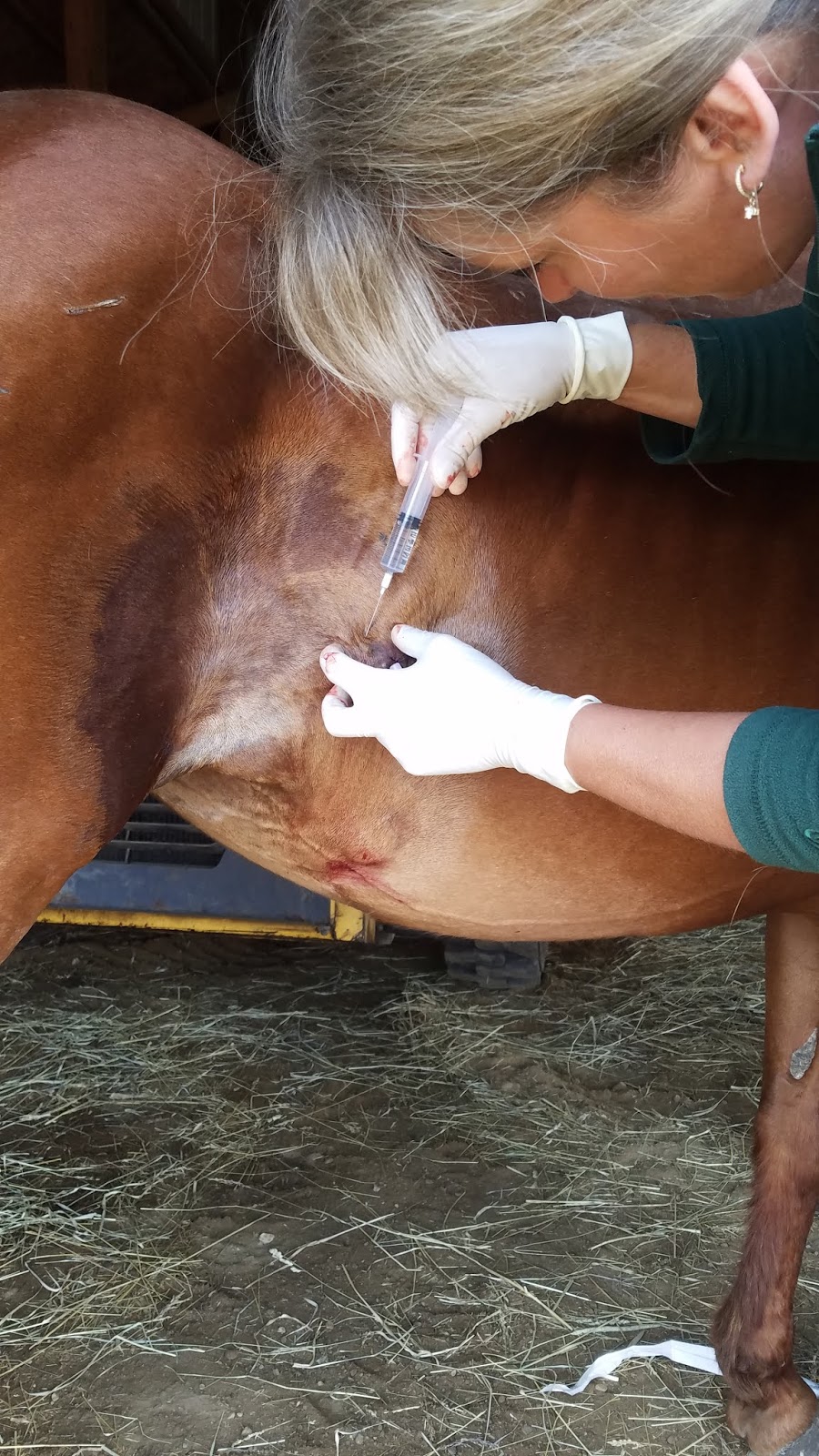
When you hear the word lymphoma you immediately think the worst, as we did at first.  But, after substantial research we found a few recorded cases of cutaneous lymphoma in horses that received treatment.  The owner did not express interest in treating the horse with chemotherapy, the most common treatment for lymphoma.  With the horses age and condition we agreed that this wasn't a feasible option.  Instead, we found positive evidence of remission with intralesional steroid treatment and oral administration of progesterone.  As this treatment was cost effective and easy to execute (the steroids were a one time treatment and the progesterone is given once daily on top of grain) we chose to give it a shot.  So we visited again, took a punch biopsy, and administered the steroids diffusely around the tumor areas.  That evening the owner messaged to say that the areas had softened up throughout the day and seemed to be getting smaller already.  We are awaiting biopsy results and typing of the lymphoma, but for now we are optimistic that we can keep the mare comfortable for a good long while.

Injecting dexamethasone into region


It is hard to believe that I am heading into my last week with Clinton Vet.  I have seen so many unique cases and met some fantastic clients.  I am so fortunate to have gotten this opportunity and I cannot wait to see what my final days have in store.  Thank you for following along!


Tuesday, June 26, 2018

Cleveland Equine Week 6

Finishing last week marked my half way point here at Cleveland Equine. It's amazing to think abut how much I've learned, and how much more comfortable I am with performing a lot of tasks.

Beanie, the mare with a corneal ulcer, is still in the hospital. I've been giving her nighttime  and weekend treatments through her subpalbepral lavage. We've adjusted her treatment plain to include fluconazole because the culture from her eye came back showing a fungal component. we also replaced the acetylcysteine with EDTA in hopes of better control the melting of the ulcer.

A barn I've been to a few times now is part of a case a couple of the doctors have been discussing. Originally, one out of three horses in the barn had a swollen eye. The vet determined the symptoms to be allergy related and dispensed some dexamethasone and suggested getting generic Zyrtec (cetirizine). We went back a second time because another horse in the barn developed the same symptoms, and then the last horse did as well. We looked for an environmental factor (they had used a new chemical in the pond that spring to kill algae, a raccoon had been hanging around the barn and there were lots of geese just outside the barn) but couldn't come up with a definitive answer. We flushed the tear ducts for all three of the horses and all are doing better on aspirin and the cetirizine.

We did another castration this week and this time I got to use the emasculators! After the vet separated the muscle from the cord and ductus deferens I first crushed/cut the muscle and then then the cord. It took more force than I thought it would! As usual, the horse was sedated with dormosedan and torb, blocked with carbocaine, and given banamine and the antibiotic genacin IV.

I also got to see two umbilical hernia surgeries this week. The first one I just watched, but for the second they allowed me to complete some of the skin sutures to close the incision! We used simple interrupted to close. The doctors commented that, while simple continuous is faster, it's riskier because if one area comes undone the entire suture line can pull out. I also learned about the vest over pants suture used during the surgery and got to practice it afterward.

The last case I'll mention here is a respiratory workup that we originally saw on the barn. The owner complained that her horse seemed slow and lazy and was having a hard time breathing during exercise. She opted to come in to the hospital a few days later for thoracic radiographs, a thoracic ultrasound and a bronchioalveolar lavage (BAL). The radiographs and ultrasound appeared normal, and we did the BAL last. This consisted of placing a tube down the trachea into the horses lungs and then flushing sterile saline into the tube and immediately pulling it back. The first sample was surprisingly clear, but the second was much cloudier and had some debris. We've sent these to be analyzed for respiratory diseases like EHV-5. I will keep you updated on what is found!

Lastly, Annie, the pony with hemothorax, has gone home! The coagulants she was given helped to stabilize her, and while she is not cured from whatever caused the bleeding into her chest, she was stable enough after about a week to return home and spend some time with her family.

I am excited for the second half of my time at CEC :)

Sunday, June 24, 2018

Cleveland Equine Week 5


This week has involved some very interesting cases! A pony has been in the ICU for four days with a hemothorax. She presented with a fever of four days duration, and we drained frank blood from both sides of her chest and ran it through the CBC. A thoracic ultrasound revealed large amounts of fluid in the chest and what appeared to be large, thickened vessels. The decision was made not to place chest tubes, as she was likely actively bleeding into her chest, and a more in-depth ultrasound was performed the next morning. The diaphragm was thickened and there appeared to be an abnormal mass very near the heart, supporting the clinicians’ suspicions of a possible hemangiosarcoma. Diaphragmatic hernia was also suspected, but no evidence of a diaphragm tear or abdominal organs in the thorax was noted on the ultrasound. Thoracic radiographs were also taken but provided little additional information because of all the fluid blocking detail.
The pony’s heart and respiration rates were consistently high, but did decrease to near normal after the first night when she received coagulant drugs that the vets normally used for EIPH horses on the racetrack. At this point the owners have given permission to euthanize when ready, but as long as she remains stable we are waiting. Beginning today (day 4) we have noted that she has become polyuric polydipsic, but continues to have a strong appetite.

Another emergency, this time treated on the farm, was a fractured sinus. The horse either hit its head or was kicked while in the pasture and had a wound between the eyes and a steady, bilateral nose bleed. Due to extensive swelling, an endoscopic exam was not performed, as significant results would have likely been obscured by blood and edema. Radiographs revealed a depression fracture. While it could be surgically repaired, the fracture did not appear to impede breathing, and the owners opted to let the fracture heal on it own, pending any significant changes in the horse’s mentation or ability to breath. Dexamethasone, bute, banamine and DMSO were all given to reduce swelling that was near the brain along with vasoconstrictors and pro-coagulants. 

One of the weeks most satisfying calls was a “lump check.” The owners had recently moved from California and wanted to establish a relationship with a vet and also have us check a 5 year old lump behind the point of the elbow that had recently tripled in size. The vet did not recommend removing it because it was not currently painful or impeding movement (it was about the size of a golf ball) and the location would require suturing in an area of high tension. The cytology sample was chunky looking, not oily like would expect from a lipoma. The cytology results plus a soft spot lead to an ultrasounded of the lump under the suspicion that it could be an abscess. Although the ultrasound did not show fluid the doctor decided to puncture the soft spot with a needle and sure enough it was an abscess! The discharge was old pus that had been walled off. Because there was such a thick capsule, the drained sac was flushed with saline and betadine to keep it open until the owner returned from a vacation the were leaving for and could necrose the capsule by flushing with formalin.
This weekend I have been giving treatments via a subpalpebral lavage that was placed Saturday afternoon. The horse has a corneal ulcer which is at risk of melting. We are waiting to hear back on culture results, but started treatments of ofloxacin, cefazolin and serum on day 1 and added IV banamine and acetylcysteine today.

Those were my favorite cases of the week; the variety of cases that I have gotten to see is great!

Brown Equine Hospital: Week 5

I apologize for the lateness of this post! It has been a busy week for me, yet again (there seems to be a theme here..) This week comprised of mainly appointments and patient care, but I will try and provide the highlights!

A horse presented to the clinic for dental work. The horse was 20+ years old and had a history of quidding. Quidding is when horses chew or ball up their hay in their mouths and fail to swallow it. This can lead to the formation of hay balls in their stalls, these are referred to as quids. Quidding can be a result of the inability to chew or swallow their food. This can be due to dental issues. In this horses’ case, they had little to no grinding surface on their molars, had molar 308 missing and some sharp edges on their teeth that needed work. Horses teeth are numbered based on location (see the attached photo).  


(photo credit: https://en.wikipedia.org/wiki/Modified_triadan_system ) 


With molar 308 missing, there was no tooth to oppose the maxillary molar (208). This is important in horses because their teeth constantly erupt throughout their lives. Their upper and lower molars will oppose one another and naturally ware each other down through chewing. With 308 missing, 208 grew unopposed and caused some issues with chewing and helped contribute to the horses quidding. With dental care, it can be addressed over time. Quidding can be an issue because it can lead to body condition changes due to lack of nutrition. This can be mitigated by adding a complete feed to the horses diet. While the horse was getting its teeth worked on, the owners wanted the veterinarian to look at a mass on the right inguinal area of the horse. While the veterinarian was palpating it, she put an ultrasound probe on there to see if it was heavily vascularized and to determine if the horse was a surgical candidate. After she was done ultrasounding the mass, she determined that the horse was a surgical candidate and that there may be more than one mass. The horse was sent to surgery the following day. The surgeon successfully removed the mass and identified four more additional masses. We took some samples of the masses and sent them off for further identification. We have yet to hear back on the results and I will try to post them as soon as I find out! Here are some before and after pictures so you can see for yourselves!

Before: 

After:


Another highlight of my week was a castration. There was nothing unusual to note about this castration besides the fact that I got to scrub in! I was so excited. The technicians taught me how to put on my surgical gloves sterilely. Once I had those on I was able to assist the surgeon. I served as the surgical technician by handing the surgeon the tools they needed and assisted during the surgery when needed as well. I was also able to use the emasculators! Emasculators are a common tool used during castrations to remove the testicles. There are two sides to these tools, a cutting side and a crushing side, therefore, it is important to ensure that you place this tool properly. You want to ensure that the crushing side faces away from the testicle (toward the body wall) while the cutting side faces the testicle. This helps to both crush and cut the spermatic chord, arteries and veins that supply the testicles. The crushing action is important because it helps to occlude the spermatic artery and prevents life-threatening blood loss. The surgeon told me that you normally want to hold the emasculators closed (tightly!) for about 2-3 minutes. Once this is done, you can use a Kelly hemostat to hold onto the remaining tissue to check for any bleeders once the emasculators are released. If there is no bleeding present, you can release the tissue. The horse recovered wonderfully and was discharged home two days later. 


The rest of my week consisted of me caring for inpatients. I was responsible for their 8 a.m. and 2 p.m. medications, TPR, feeding, watering, haying and picking stalls. The more time I spend in the hospital the more responsibilities I am given. I am really enjoying the responsibilities that I am given and I enjoy the challenge. I am getting much better with placing catheters, which is great! I have a tendency to rely too heavily on my safety blanket (the techs!) and they are slowly taking it away from me, which is good. This allows me to depend on myself and my intuition, which I am learning to trust. 

We will see what this next week brings me! Good night!


Littleton Equine Weeks 6 and 7

Hello everyone! Apologies in advance for the brevity of this post- my computer has been on the fritz the past two weeks, but I had it taken care of yesterday.

LEqMC has been busy as ever! This past week was bittersweet as the 2017-2018 interns finished their last day on Thursday 6/26 and the new group of interns started. Since the last time I wrote, young Pheonix has returned home! He had two procedures to fix a stricture of his rectum so he can defecate normally. The poor guy has to be bedded on grass hay because he eats all other types of bedding.... (shavings, straw, and we certainly don't want to put him on sand!) Luke, the young Shire (I thought he was a Percheron, but I was wrong) ended up going to colic surgery. I was unable to witness it, but he has since gone home and is on the mend!

I've seen lots of lameness exams the past two weeks, and it has been really fun refining my skills in noting where the lameness is. I've also seen lots of joint injections, which is helpful for remembering important equine anatomy.

There have been SO MANY colic cases here, it seems that it's really common here in Colorado. One of the cases this past week was an American Cream draft who came in for sand colic. He was medically managed for 4-5 days before it was decided that he would go to surgery as he was not improving and not passing the sand. Often pain levels are a good indication of how bad the colic is. It's difficult to tell how sick a draft horse is based on pain because they are so stoic. There is concern with taking sand colic cases to surgery because the tissue can weaken and become friable, leaving the horse more susceptible to rupture when the large colon is exteriorized during surgery. Luckily, Hickock's surgery went well after removal of around 80lbs of sand/gravel, feed material, and mineral oil. There was some concern about the integrity of a few areas of large colon that appeared weakened. We're now 4 days post-op and he's still doing well. He's eating and going for handwalks now.

During my ICU days this past week I had the chance to spend some time with Dr. Duff, watching pregnancy checks. I watched him lavage for an embryo and had the opportunity to look for it under the microscope. Unfortunately, we didn't get it but it was cool to practice looking and learn about embryo collection.

Last Friday I was able to ride with Dr. Dunbar, who has a special interest in dentistry. I watched her perform several floats and dental exams. We discussed the importance of having a speculum, excellent light source, sedation, and a mirror in order to do a proper dental exam and float. One of her patients was a Belgian mare with sidewinder. She did not have her teeth floated but was vaccinated and examined. Having sidewinder put that mare at risk of falling under sedation, risking injury or inability to rise once the sedation wore off.

This past Friday I went to the horse show with Dr. Tisher and Dr. Pierson. I jogged horses for lameness exams and restrained for pro-stride treatment and two schockwave treatments. I really enjoy equine sports med, and you definitely have to have a well-trained eye for lameness as some of them can be subtle (but important to their athletic performance).

Friday, June 22, 2018

Equine Athlete: Week 5-6

Hello from Wilmington Ohio. Equine Athlete will be on the road for the next couple weeks. We arrived here on Monday for the AHA Region 13 Championships. Next Monday we will be headed to Lexington Kentucky for Region 14 Championships. With that, I’d like to fill you in on what a day at the shows consists of. We arrive to the facilities as everyone is hauling in for the week, in case of any travelling issues. There have been a few the last couple of days, just some minor colics, which we have treated with fluids, and electrolytes. Aside from the general care we do various treatments related to sports medicine. We provide shockwave therapy, therapeutic ultrasound, laser therapy, chiropractic adjustments, and much more.

Horse show life consists of early mornings and late nights. The different therapies that I mentioned above have to be perfectly timed out from when the horse shows. Therefore our schedule is pretty jam packed. Aside from our set schedule, we get numerous of calls for unexpected problems that we must resolve. If a lameness issue is presented we first observe if it is a three leg lameness or something less obvious. If it is a three leg lameness, there are usually four possibilities; a fracture, foreign object, abscess, or the horse is septic. We will access these horses for any wounds and palpate to find where the issue is stemming from. The majority of three leg lameness’s that I have seen this summer are from an abscess and we use hoof testers and x-rays to determine if that is the problem. If is it something less obvious, such as a suspensory or a joint issue, we will lounge the horse first followed by flexing. Flexing is a stress test used to pin point where the pain is coming from. Lastly, palpations are done to reassure the findings on the lounge line and flex test. Using what was found a treatment plan is put into place, it is always different depending on the individual. At the shows we can’t do joint injections because it have to be 7 days from when the horse shows, but we can use one of the other therapies listed above in order to make the horse more comfortable.

With that, being at a horse show is a lot of running around, we rarely get any breaks. We have been working on average 15 hour days so it can be exhausting but I am loving every minute of it and I’m learning so much!


I hope to see some more interesting cases the next couple of weeks that I will share! Stay tuned!