Wednesday, July 31, 2013

Farewell to Somerset

Well, it has finally sunk in that my time with the MSU Equine Summer Fellowship Program and Brown Equine Hospital has come to an end. I could not have imagined a better experience and I would do it all over again in a heartbeat. The vets and techs at Brown Equine Hospital taught me so much and were so supportive, I could not ask for a better group of people to work with. I was very sad to say goodbye, but I left with many fond memories. So here it is, my final blog post:
Something must have been in the air this week, because we received four emergency colic cases in less than three days. The first to be brought in was a draft horse that had been off feed since the night before. From the abdominal ultrasound and rectal exam, Dr. Brown diagnosed him with anterior enteritis, or inflammation of the duodenum and/or jejunum. Since anesthetizing draft horses carries an even higher risk than other horses, Dr. Brown wanted to keep this gelding off the table at all costs. We started by passing a nasogastric tube to reflux every few hours and administering IV fluids with a lidocaine drip. The draft horse took a turn for the worst a couple days into treatment, refluxing up to 30 liters and going into acute renal failure. We increased the frequency of refluxing and started to bolus the IV fluids. Remarkably, the gelding pulled through and is now recovering well. We stopped refluxing completely and have started weaning him back onto solid food.
The second colic that came in was a part-Standardbred that had been acting uncomfortable for a couple days. As it turned out, this gelding also had anterior enteritis and we started him on the same treatment regimen as the draft horse. Unfortunately, our refluxing did not keep pace with the fluid backing up into his stomach. About 48 hours after being admitted, we passed a tube, but we got negative net reflux. Suspicious, Dr. Brown performed another ultrasound and belly tap. The results showed excessive fluid (reflux) surrounding the intestines, revealing that the gelding’s stomach had ruptured. Sadly, we had to put the horse down. It is surprising how such similar cases, treated the same, can end so differently.
The last two cases were also treated medically. An impaction and a right dorsal displacement were resolved with IV fluids and fasting. Both horses were slowly reintroduced to solid food and were sent home within two days of being admitted. I had always thought that all colic cases that were referred went to surgery. Much to my surprise, however, the vast majority of the colic cases we saw this summer were treated and resolved medically. Another surprise was how many of the horses that came in for colic went home healthy; it was nice to discover that colic is not a death sentence.
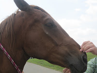
To end, here are a few photos of some of my favorite moments working at Brown Equine Hospital:
Repro work with Dr. Jen Brown

Surgery with Dr. Keith Brown

Scoping with Dr. Travis Tull

Sunday, July 21, 2013

Week Nine in Somerset

It is so hard to believe that I will leave Brown Equine Hospital in less than a week. This summer has flown by and I have learned so much in the short time I have spent here. Like the summer, this past week sped by and we had a full caseload of horses to be scoped.
The most invasive procedure we performed with the scope was a ventriculocordectomy. A ventriculocordectomy is the removal of the laryngeal ventricles and vocal cords to enlarge the airway. During this procedure, the scope is passed through the nose to the level of the epiglottis and gives the veterinarian a clear view of the larynx as he works. The ventricles and vocal cords can then be removed through an incision in the throatlatch area. In our first case, the patient had partial paralysis of the left recurrent laryngeal nerve, which prevented him from fully opening his left arytenoid cartilage and vocal cord. Dr. Brown is confident that the ventriculocordectomy will allow him to perform at a high athletic level despite the partial paralysis.
We suspected that a second horse presenting with exercise intolerance also had laryngeal nerve paralysis. This gelding had an even more pronounced paralysis in both the left and right arytenoid cartilages and the owner was already considering a ventriculocordectomy. Upon close examination of the horse as a whole, however, severe facial muscle atrophy on the right side of the face was also observed. Dr. Brown diagnosed the horse with Equine Protozoal Myeloencephalitis (EPM), a common neurological disease. Instead of staying for surgery, the gelding went home to be treated with anti-protozoal drugs and a vitamin E supplement.
Minor muscle atrophy of the left side of the face compared to advanced muscule atrophy of the right ride

We used a gastroscope to go up the nose, under the epiglottis, down the esophagus, and into the stomach of another patient to check for gastric ulcers. Surprisingly, once we entered the stomach, we found no ulcers. Instead we found clusters of bot fly eggs (truly disgusting.) Our final scope case was the guttural pouch fungal infection that we have been treating topically through the scope for the past three weeks. We are now able to tear pieces off of the fungal plaque and inject the topical treatment directly into it. Below is a series of pictures documenting our progress.
    
The plaque upon presentation, during a lavage with dilute betadine, after two weeks of treatment

Topical treatment with an anti-fungal suspension, after three weeks of topical treatment

Despite being busy, we still found some time to have fun. One of Dr. Brown’s heavy weight pulling horses, Tank, has been staying at the clinic so Dr. Brown can work with him.  The externs and I go along for the ride when we can to add weight to the sled; we have a grand time trying to keep our balance on the moving pallet. Dr. Brown even let me try my hand driving Tank. It is never a dull day around here!
        

Thursday, July 18, 2013

Time Flies When You’re Working Hard, Having Fun, and Learning Loads!


Time is flying by here at CEC! I am just finishing off my tenth week and I am still having an awesome time! I have seen so many neat cases and am learning so much. Even though, it is in the 90s this week, I love working hard and going on lots of farm calls. This week we had an interesting wound that was along the lateral canthus of the eye. Somehow the horse had avoided scratching the eye and we were able to suture the wound closed with 3-0 vicryl.  The real test will be whether the owners can stop the horse from rubbing the eye, which for this trouble-maker of a horse will be a challenge!

There have been several colic cases lately. One involved an emergency farm call out to see a draft horse 5yr mare. The mare had a heart rate of 100beats/min, respiratory rate of 80bpm, temperature of 103*F, muddy-color gums, and a severe sweat. I was able to rectal the mare and felt an extreme amount of heat, several distended loops of small intestine, and a very firm cecum. Unfortunately, because surgery was not an option for this mare, she was euthanized. We have also been seeing several impaction cases, ranging from mild to severe. We currently have an impaction colic case that has now developed severe diarrhea and a fever. A fecal sample has been submitted so we will see what it says, however I’ve been told that it “smells like Salmonella!” The horse is now on K-pen, gent, polymyxin, cimetidine, and fluids. In addition, to continuing multiple day NG tubing with different combinations of water, electrolytes, energy, Epsom salts, and mineral oil. I’ve found colic cases to be very interesting, although often frustrating (if only you could make a horse drink)!

I can’t believe that I only have two week left. I truly can’t say how much I love working here!

Wednesday, July 17, 2013

Hard Lessons from RREH

Hello again from the Bluegrass! As summer winds down here, I thought I’d take a moment to talk about some of the hard lessons that we learn in equine/veterinary medicine.

Yesterday started out as a pretty routine day for surgeries with Dr. Bramlage, but right around 1:00 PM, we had an emergency that came in – an internal carotid hemorrhage in the right guttural pouch… and when I say hemorrhage, I mean pouring out! This mare was a 12 y.o. TB mare that had actually been in sometime between late April and early May with a similar presentation, and had actually had her internal carotid already ligated surgically.

Initially when she came in, she left a trail of blood from the parking lot to a pool in the work-up area and induction stall. Prior to and during surgery, she had probably lost approximately 20% of her blood volume (so roughly about 10L – horses have a lot more blood than the average human). During surgery, she had part of her volume replaced with fluids, Hetastarch, and plasma, and she was eventually matched with one of the blood donor horses for a transfusion following surgery. The surgeon on the case performed a balloon embolization to stop the bleeding from that artery – which, essentially, puts a “plug” in the vessel by inflating the end of a small, wire-like catheter with saline (almost like filling a miniature water balloon that blocks the blood flow).

The surgery was successful at stopping the hemorrhage, and the mare was placed in the recovery stall and fitted with a blood transfusion set. Two of the best recovery guys were with her the entire time, and were extremely conscientious of her condition and how she was progressing. It took a good hour and a half for her to regain enough strength and come around from anesthesia to even try to stand up with their assistance – not entirely unexpected due to how she came in and how long the surgery took (about 3 hours!). The first time, she couldn’t stand up fully, and the team allowed her to rest in lateral, a.k.a. on her side, a little bit before trying to help her up again. She was still incredibly weak from all of the fluid she lost and probably some electrolyte imbalances, etc.

Once she sat up sternal (a.k.a. on her belly), the guys and an anesthetist adjusted her hooves so that if she tried to stand again, she would be in a better position for success. Again, she tried to stand with their assistance, but, she was still too weak and collapsed from fatigue – this time, it was not a smooth fall. To everyone’s dismay, when she collapsed, she also completely fractured her right tibia (for all you basketball fans, think Kevin Ware in the NCAA tourney). Tibial fractures in horses are basically fatal because they cannot be easily repaired without major complications. And, fractures during recovery are every veterinarian’s worst nightmare.

When this happened, the interns sprung to action and were able to sedate her until her owners could be reached, and quite regrettably, she was euthanized. No one was truly to blame in this situation, nor did anyone expect for this to happen. It’s just one of the unfortunate and undesirable things about equine surgery. Mostly, I feel for the surgeon and the intern on her case – it’s devastating to complete a difficult, 3-hour-long surgery and have a positive outlook for your patient, and then have something so tragic happen that absolutely no one could control.

So, this blog is in memory of the sweet mare that we lost, though please do not let it scare you – fractures during recovery are rare if the recovery team is good at what they do, and they should never be anticipated as though it will inevitably happen. Props to our recovery guys, who watch out for the safety of all surgery patients each day, and even risk their own safety for many of our horses!

Monday, July 15, 2013

Week Eight in Somerset


We started the week off with our normal cases of joint injections, lameness exams, pregnancy checks, and various other procedures. Although I have not yet discussed it, seemingly every week at least one overweight horse comes in lame with some degree of laminitis and rotation. Equine Metabolic Syndrome is something that I had vaguely heard of before, but I have gained a much better understanding of the prevalence of the problem here. EMS is developed insulin resistance comparable to Type II diabetes in humans. For some reason, changes in the sugar composition in the grass during spring and fall cause insulin resistant horses to experience bouts of laminitis. These horses also become very overweight, depositing fat around their tail-heads and developing a “cresty” neck appearance. We have seen so many cases now that I can tell by looking at these horses that they will have coffin bone rotation on a radiograph. Just like with diabetes, the solution to the problem is diet and exercise. Dr. Brown and Dr. Tull recommend switching to a low-starch grain, severely limiting access to grass, and soaking the hay before feeding to leach out the sugars. EMS is an interesting condition and I am sure I will be seeing it quite often, even after I leave Somerset.
On Tuesday, we had the surprise of not one, but two horse with P1 fractures come in. Both cases were lateral condylar factures that were repaired using two lag screws. As always, I got to scrub in! It was a long day in the operating room, but Dr. Brown set both fractures with relative easy. I finally got the hang of assisting for these fracture repairs (at first all of the hardware and new instruments intimidated me) and could anticipate what Dr. Brown would need next. In the end, he let me practice my vertical mattress suture pattern to close over the screws. We were all pretty excited over how well the surgeries went. Both horses recovered well and were sent home a few days later. 
The post-op radiographs. You can't even see the fracture line!

   
Drilling                                  Flushing                                Sewing

The twist of the week came on Wednesday, when I started experiencing severe abdominal pain and my Brown Equine family decided I needed to go to the ER. After every diagnostic test under the sun, the human doctors admitted me to the hospital for several days on fluids and pain meds. The final diagnosis was a viral infection and they released me after I had no pain or fever for 24 hours. While the nurses and staff were very nice, I am more than excited to be back at work and I am looking forward to another great week.

Thursday, July 11, 2013

Weird things at RREH

Hey everyone! I hope your summers are treating you well. We’ve enjoyed some warm and sunny weather here in Lexington, with the occasional rainstorms, of course. I wanted to take this week’s post to discuss the “weird” cases that we’ve seen. The majority of these cases have come in as emergency rather than elective cases, and unfortunately the outcomes aren’t always what we hope for.


  1. TB broodmare with a foal at her side presenting for a moderately painful colic:

On evaluation, nothing major is noted about her large colon (wall thickness, etc), and for the most part, the majority of her small intestine was just moderately dilated. Regardless, she was taken to surgery as she was insured and of significant value to the farm. The surgeon on her case, Dr. Embertson, found that (oddly enough) she had a moderately large diaphragmatic hernia, with protruding small intestine and the cranial portion of her spleen… quite a rare finding! The intern on the case even noted that she was able to reach through the diaphragm and feel the heart beating – which, in and of itself, was impressive. Normal repair of diaphragmatic hernias in horses often require implantation with a prosthetic mesh across the lesion, but RREH rarely keeps mesh on hand because of how rarely it is used. Dr. E repaired it by repositioning of the viscera and careful appositional suturing, and to date, the mare (with her foal) is doing well in recovery!


  1. Emergency presentation of a Quarter horse gelding (yes, I did say quarter horse!), with a severe right hindlimb lameness after showing:

This gelding came in after hours on the weekend after barrel racing, and was essentially 3-legged lame. Upon radiographs, it was discovered that he had a severely communited P1 fracture. In fact, I would not even call it a fracture – I would call it a shatter. There was hardly any intact bone that could be used as a “strut” to help reconstruct the pastern, to at least allow him to live as a retired pet. He had two options: cast the limb and see how it healed on its own, or euthanasia. In all honesty, casting the limb had an extremely poor prognosis for any return to function, and it was suspected that he would develop other complications during the healing process, such as laminitis in the contralateral limb, etc. The owner reluctantly and difficultly chose to euthanize him, and we all felt for her and her situation. I can’t imagine what I would do in her situation (knock on wood that it never happens to anyone…).


  1. Young TB mare presenting for an elective fetlock arthrodesis:

This mare presented as a Dr. Larry Bramlage case (the master of most things orthopedic) for front fetlock (cannon bone to phalanx 1) arthodesis, mostly as a salvage procedure. I have personally experienced Dr. Bramlage’s mastery of joints and bony structures, including one fetlock arthroscopy lasting merely 5 minutes… but even this surgery was a challenging one and it took several hours. Dr. Bramlage used a standard method of LCP plating on the dorsal surface of the bones with 4.5 and 5.5 mm screws (in this case, he used a 12-hole plate). Again, this is entirely a salvage procedure to allow the mare to be more comfortable in her fetlock in her future years, although the procedure itself does create quite a substantial amount of pain in the initial recovery period. However, like many of our orthopedic cases, she is recovering well here in the hospital and is regularly having her cast changed to prevent pressure sores.


Like most of the other fellows, I try to take the free time that I have to enjoy the culture and city of Lexington, and the different things it has to offer. There’s a lot of history here, and in my opinion, some really great “foodie” places! Here are pictures of some of my favorites thus far:

Stella's Kentucky Deli: a to-die-for homestyle brunch place with all you can eat cheese grits, stratas, orange pecan pancakes... yum. Definitely give it a try if you are in town!

The Local Taco: some of the best Mexican food I've had anywhere. The mole sauce is particularly delicious, as is the sangria, if you are old enough!

North Lime Coffee and Donuts: a quirkly little place featuring delectable espresso and tea drinks, as well as fun doughnut flavors like Root Beer Float, Strawberry Yeti, Chocolate Covered Blueberry, and Pink Lemonade, to name just a few!


Did you know that Lexington is actually the childhood home of Mary Todd Lincoln, wife of President Abraham Lincoln?! This is her historic house, which sits right on Main Street in downtown Lexington.
 

Sunday, July 7, 2013

Week Seven in Somerset

We celebrated the Fourth of July this week by welcoming two overseas vet students to the clinic. Jenny is originally from West Virginia and is going to school in New Zealand; Sonea is from Austria and is going to school in Vienna. It is really interesting to hear about school and life in general from a foreign perspective. As I show them the ropes around the clinic, they are sharing their knowledge about the cases with me.

This week was full of new cases, exciting hands-on experiences, and very little sleep. We had two yearling Standardbreds in for arthroscopies to remove OCD lesions in their hocks. Assisting for these arthroscopies has become less daunting for me, despite all the strange instruments. I am also finally able to identify anatomical landmarks on the screen. I was pumped beyond pumped when Dr. Brown allowed me to remove OCD lesions from the distal intermediate ridge of the tibia in both hocks for one of the horses. I have a new appreciation for the coordination required to grasp a small chip with only the small view on a computer screen for guidance. Dr. Brown makes it look easy. The second arthroscopy revealed that the chip we could see easily on an x-ray was actually buried in a collateral ligament. Dr. Brown and Dr. Tull were both very excited to examine the soft tissue structures of the joint with the scope. They figure that the horse tore his deep short medial collateral ligament, which carried a chip from the medial malleolus with it. Dr. Brown was able to clean up the torn ligament and remove the chip, but he does not hold much hope for the horse’s racing career.
We also had two more foals with swollen joints come in, but neither of them turned out to have septic joints. The first has an infected physis, or growth plate, which we are treating with systemic antibiotics and regional limb perfusions. The second, Dr. Tull suspects has Rhodococcus, based on his findings from a thoracic ultrasound. Rhodococcus is a bacterium that can cause pneumonia in foals by forming abscesses in the thorax. Due to the connect blood supply between the metaphysic and epiphysis in foals the bacteria can gain access to the joint area, causing inflammation around the joint. For this reason, sometimes the most visible sign of Rhodococcus is swollen joints. We are treating the infection with targeted oral drugs that are proven to penetrate the abscesses.

        
Here are some action shots of the arthroscopy surgeries. On the far left, I am "assisting" in a whole new way. In the middle two frame, I am examing and gripping two OCD lesions on the computer screen in preparation of removal. The final picture is of my proud display of the first chip I removed. 

Tuesday, July 2, 2013

Update from St Johns


It has been a few weeks since my last blog entry. I have been busy working for MSU’s advising office at the Academic Orientation Program (AOP) for incoming undergraduate freshmen. This is my second summer working the program and I present the pre-veterinary and veterinary technology programs to the undergrads who have declared their major as such. Due to my prior commitment with AOP for six weeks this summer, I am only able to be with Clinton Veterinary Service on Fridays until AOP is finished.

I am incredibly thankful to Clinton Vet for being so accommodating and flexible with me for these next few weeks. I have still been able to log a few hours every Friday still assisting with checking breeding cycles and pregnancies at MSU’s horse farm, various teeth floats, changing bandages, and assorted other appointments. I look forward to being back with the clinic full time in a few weeks!