Saturday, May 31, 2014

Weeks 3 and 4 at Brown Equine Hospital

Weeks 3 and 4 at BEG have been relatively slow compared to my first 2 weeks, but we still have plenty to do.  Dr. Jen Brown has had a steady flow of mares in and out of the clinic for breeding services so I have gotten the chance to do some rectal palpations and assist with artificial inseminations.  A lot of this reproductive work is familiar to me due to my time working at the UC Davis Horse Barn in undergrad, but it is always nice to brush up on these skills.

There have been a variety of cases over the last couple weeks, but I will focus on a couple in this post.

One case was a horse that had an episode of colic a few days prior, and the owners had given Banamine® (Flunixin meglumine, a common NSAID used in horses and cattle) in the muscle.  While this drug is labeled for intramuscular injection, there is a risk of the horse developing a serious condition called Clostridial myositis.  This occurs when Clostridium bacteria dormant in the muscle or on the skin surface proliferates in the muscle producing gas under the skin and release toxins into the blood stream.  This condition is serious and can be fatal.  In addition to giving systemic antibiotics, the method to treat Clostridial myositis involves making incisions into the infected muscle to clean and expose the bacteria to air (Clostridium is an anaerobic bacteria, and does not like being exposed to air).  Unfortunately, the owners elected to have the horse euthanized.  Many people, even some vets, give banamine (and other drugs) in the muscle and never experience this devastating disease, but clients should always be educated about the risk associated with intramuscular injections of certain drugs.

Another case that we are currently working on involves a horse that had a tail wrap applied too tight causing the tissue around the tail to die.  The referring vets tried to remove the dead parts of the tail in addition to several vertebrae, but were unable to close the wound fully.  Warning: The following images may be a bit graphic for some people. 

The tail was very short on presentation to Brown Equine Hospital.  There was some dirt and various topical antiseptic agents on the open wound so we clipped and cleaned the tail and wound to better assess the injury


After cleaning, we could see the wound was a mixture of necrotic (the black tissue) and granulation tissue (the bright red tissue).  


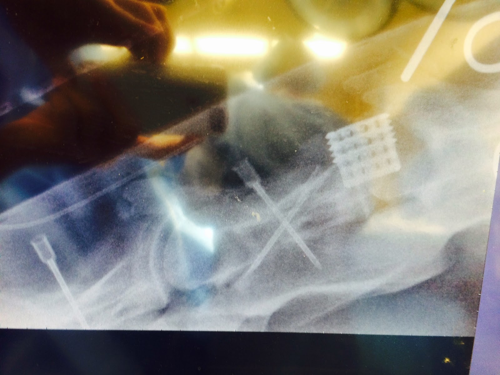
A radiograph of the tail prior to surgery
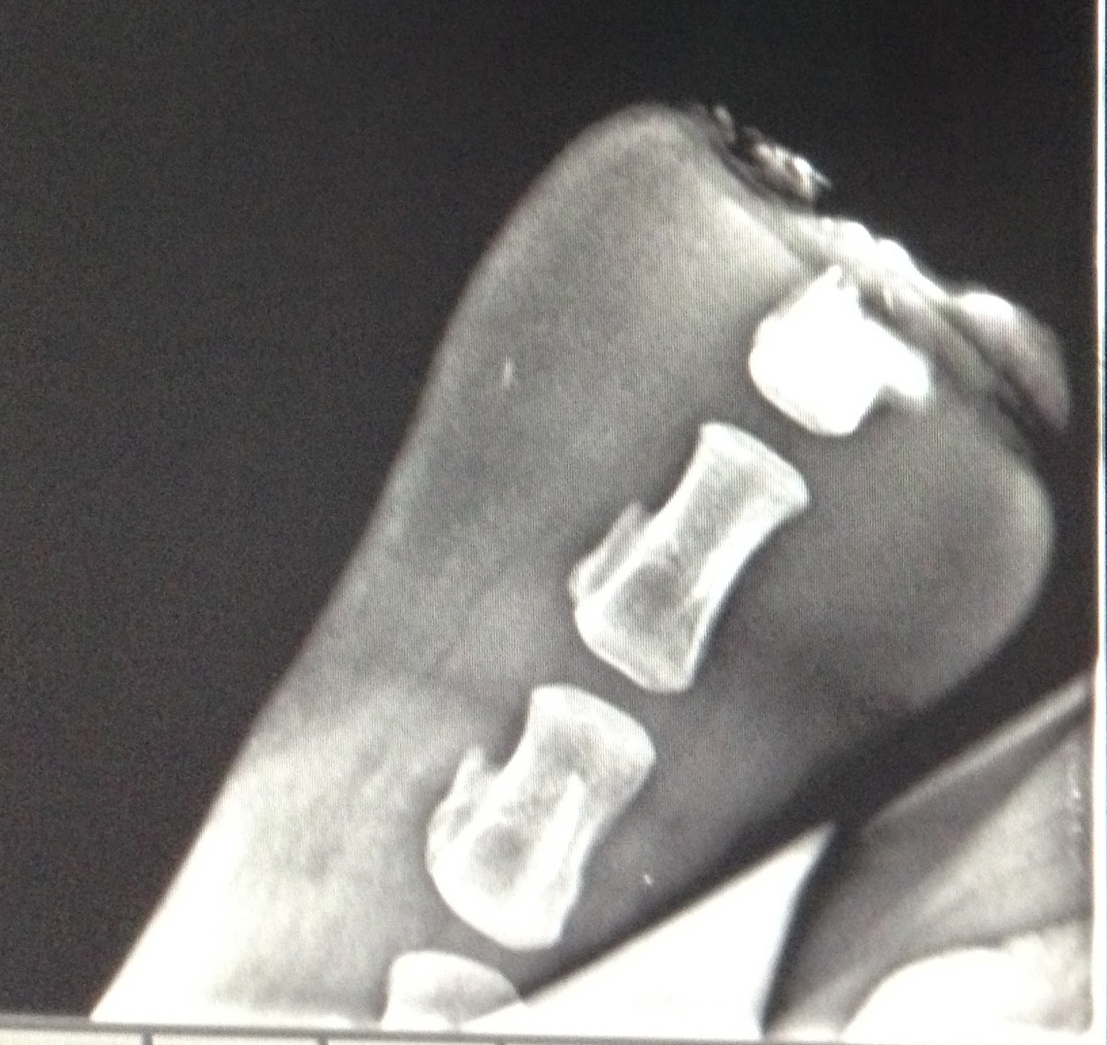
At the tip of the tail, there was part of a vertebrae remaining from the prior amputation attempts.

Dr. Hackett and Dr. Younkin removed the remainder of that vertebrae and a great deal of the granulation tissue in order to have enough skin to close over the end of the tail.


Closure with a Near-Far-Far-Near suture for tension relief, and then a simple interrupted pattern was placed in between the tension relieving sutures for apposition of the raw edges.

The patient will need to be on systemic antibiotics and anti-inflammatory drugs for several days to prevent further infection and minimize pain, but the prognosis for recovery is good!

The next couple weeks will bring several big changes to the clinic.  Dr. Younkin will be finishing his internship at BEH and heading off to Kansas State for a new position.  He has been a valuable source of information in my first month at BEH and we wish him only the best as he advances in his career.  A new intern who recently graduated from University of Pennsylvania will be starting next week, and BEH will also be welcoming a new board certified surgeon to the team from Tufts University, Dr. Patricia Provost.  I am looking forward to having even more people to learn from! 


Until next week!

Wednesday, May 28, 2014

Getting my feet wet at Clinton Veterinary Service: My first 2 weeks

I’m Valerie, a veterinary student from Michigan State University who recently finished second year and is looking forward to a summer of equine work. The first two weeks at Clinton Veterinary Service in St. Johns, MI flew by with remarkable speed. The practice currently has 6 doctors who largely work as a large animal ambulatory practice with some equine cases trailered in to the clinic occasionally. This summer I am primarily riding along with Dr. Cynthia Trombley, an equine vet who graduated from MSU in 1995 and co-owns the practice with her husband. From my first day onward, we’ve been quite busy. This time of year the majority of the calls are routine maintenance appointments: vaccinations, Coggins testing, health certificates for any horses traveling out of state, fecal floats and consultations on deworming regimens, and checking teeth for any needed dental work. In my short time here, the difference regular care and preventative medicine makes in the health of a horse is already readily apparent. With Michigan’s long winter, many people are just now transitioning their horses to pasture, which means we’ve had an increased number of colic calls. The majority of the cases have been gas colics, likely caused by the switch to fresh pasture, and have been successfully treated medically on farm. Fortunately few of the colic cases we’ve seen since I started needed surgical treatment, but our close proximity to MSU is helpful in the event surgery is warranted and the owners are willing to pursue that option.
                Dr. Trombley also sees a modest amount of reproductive work. Three days a week we visit a large Arabian farm to ultrasound mares. Ultrasound is useful for tracking a mare’s cycle, identifying follicles that are likely to ovulate, and confirming pregnancy. Unlike in cows, pregnancy can be diagnosed as early as 14 days by ultrasound. While I have yet to palpate or ultrasound a mare, I am becoming much more comfortable with interpreting what I see on ultrasound. The repro work is enjoyable and particularly rewarding when it results in a successful pregnancy. Since the decline of the racing industry in MI and the recession, there have been far fewer clients breeding horses these days, but we still have been out to several other farms to ultrasound mares, collect samples for uterine cultures, perform breeding soundness exams. The most gratifying calls are the new foal check up calls. I enjoy working with foals, but seeing the owner’s excitement over new life and new potential is very satisfying. Hearing owner’s talk with deep pride in their voice about the new foal that is the result of many generations of careful breeding and years of commitment to a breed or disciple is a good reminder that we as veterinarians may treat animals, but we do so in the service of people. We work to maintain the health of animals that bring many people a great deal of joy.

                I have seen and learned a great deal in the 2 short weeks here at Clinton Veterinary Service, and I’m looking forward to the weeks to come.


 A new foal visit to check out this nice little colt. It is also advisable to examine the placenta whenever possible after parturition.

Upper Respiratory Fun

            My time this past week has been divided between the road and the clinic. Until now, most of my experience has been in a hospital setting, so I have been quite impressed by the variety of diagnostics and treatments that can be provided out of the back of a truck. This week alone, I have assisted with over half a dozen stall-side respiratory endoscopies. The hand-help scope, instead of being connected to a computer monitor, has an eyepiece that allows the vet to visualize the internal structures while still controlling the camera. We used the scope to diagnose dorsal displacement of the soft palate, rule out guttural pouch infection, visualize chondritis of the arytenoid cartilage (swollen larynx), and perform alveolar lavages to test for lower respiratory infections. Most of the time the horses require little to no sedation for this procedure.

            I spent one afternoon with Dr. Berthold as he treated three horses with dorsal displacement of the soft palate, also referred to as flipping the palate. Normally, the soft palate should sit snuggly underneath the epiglottis of the larynx. Inflammation, irritation, or malformation of the epiglottis can cause it to slip underneath the soft palate. Most horses can self-correct the displacement by swallowing. In those that don’t self-correct, the soft palate becomes an airway obstruction that can affect athletic performance. Dr. Berthold takes a two-step approach to correct chronic displacement. First, he uses a laser, directed by the endoscope, to cauterize the soft palate. Ideally the resulting inflammation and scar tissue will help keep the epiglottis above the soft palate. Next, we laid each horse down to perform a myectomy. This procedure entailed making a midline incision over the cricoid cartialges and first few tracheal rings, then removing the tendonous insertion and part of the body of the sternothyroideous muscle, and finally removing part of the body of the sternohyoideus muscle. The purpose of the myectomy is to inhibit the backward motion of the epiglottis that allows it to slip under the soft palate. Dr. Berthold has gotten very good results by combining the two strategies. A huge advantage of doing this procedure in the stall is efficiency; we had three procedures completed and the horses recovered in less than three hours. The drawback is that recovery can be a little more risky. Although the horses were anesthetized for significantly less time than if done in a surgery suit, they recovered in a wooden stall instead of a padded room. It is always interesting to see the pros and cons of different approaches and compare a farm call to a hospital environment.

Monday, May 26, 2014

Week 3 @ SVEC


            My third week at Saginaw Valley Equine Clinic ended up being a bit quieter than the first 2.  The foal from last week has continued to get stronger and stronger (and getting a little feisty too) and is now completely off treatments.  We continued this week with much of the usual – vaccines, Coggins, lameness exams, joint injections, checking mares, collecting stallions, and breeding.  We did have another gastric scope, and I can now say that I am pretty good at passing the scope!  We also castrated a 6 month old colt which was a good experience for me as Dr. Jones dropped him for the procedure where I have previously only observed castrations done standing.
            Friday turned out to be a very long and sad day with 2 euthanasias.  A 26 year old horse had gotten cast in his stall and after making it back to his feet, was completely non-weight bearing on his left hind leg.  Based on how he was holding it and how his muscles looked, Dr. Jones thought he may have fractured his femur.  The owners chose to euthanize without doing any diagnostics since the eventual outcome was likely going to be the same.  The other horse was only 16 but he had foundered several years ago and their success at managing his pain was starting to dwindle.  Before Friday, I had never seen a horse euthanized and as with small animals, it was very difficult to watch as the owners clearly had a deep connection with their animal and it was hard for them to say goodbye, even though it was for all the right reasons.
            And it wouldn’t be a Friday without an emergency.  This one happened to be a recipient mare owned by the clinic.  Owners who want to breed their mare but don’t want to lose a show season due their horse being in foal often lease a recipient mare.  The mare is bred normally and then 1 week later, before the embryo implants into the uterus of the mare, the embryo is flushed out and put into the recipient.  This mare is very sick, and fortunately, is not in foal.  The barn crew thought she wasn’t quite right for a few days as she was isolating herself from the rest of the herd but by Friday she had a temperature of 104, severe edema in all legs, skin sloughing off her nose, and her conjunctiva was icteric.  She was brought into the clinic and placed in isolation.  I had the weekend off so I was not there to see how this case has progressed, but I am very curious to see how she is doing tomorrow morning and find out if they have determined the underlying cause of her condition.

Sunday, May 25, 2014

Greetings from Rood and Riddle!


      These past two weeks have been busy busy in the surgery department! 90% of the surgeries seen here are often transphyseal bridging or arthroscopies. Transphyseal bridging is done on horses that are one year old or younger to correct uneven growth of a foot and usually involves inserting a staple or series of screws and wires on the knee or fetlock to inhibit growth on one side of the joint, giving the other side a chance to catch up. Once a limb has corrected, the staple is removed. If the implants are not removed, the angular deformity will overcorrect, and the leg will acquire a deformity in the opposite direction. 
       Arthroscopic surgery is minimally invasive surgery in which an endoscope attached to a camera is inserted into the joint via a small incision. The surgeon can then view the inside of the joint on a video monitor. Because the joint does not have to be completely opened, the recovery time is usually significantly reduced, and the success rate may be greater due to a decrease in trauma to the connective tissues around the joint.  In cases of OCD or chip fractures (which is what we see almost everyday here!), the horse is placed under general anesthesia, the affected joint is prepared for surgery, and two small incisions are made in the skin overlying the joint. One incision allows placement of the arthroscope in the joint so the surgeon can visualize the lesion, and the other incision allows the instruments to be placed in the joint to allow chip fracture removal or debridement of an OD lesion. 
      On top of our usual days, there is always a special case in the mix which keeps things interesting! One interesting surgery we did was a cervical spinal cord surgery. This surgery is done on horses exhibiting Wobbler Syndrome, which refers to a number of disease states in the horse. The most common is termed cervical vertebral malformation and is characterized by malformation or compression of the spinal cord which leads to spasticity, ataxia, and incoordination.These symptoms are caused by damage to or compression on the spinal cord.  The surgical technique involves drilling a hole between the affected vertebral bodies from underneath the neck and inserting a stainless steel prosthesis called a “Bagby Basket,” which fuses and immobilizes the vertebrae.
Dr. Woodie performing the basket placement surgery 

Basket implant on a radiograph of the spinal cord


     Another interesting case this week was a mare brought in for an ovariectomy, which is a surgery that involves the removal of a diseased ovary. This is a commonly done procedure, and can be done on the farm. However this was a special case, as this ovary came out to be about the size of a bowling ball on ultrasound! Dr. Woodie had to remove about 1 liter of fluid out of the ovary before we could even think about trying to remove it out of the body. 

This image represents a normal ovary, with the one on the left being during non-breeding season, and on the right during breeding season. 

This is the ovary we removed, clearly diseased, and much larger. 

Thanks for reading our first post from Rood and Riddle!!!!

-Lisa Reznik 


Friday, May 23, 2014

Hello from Colorful Colorado

Hey everyone,

I have been working at Littleton Equine Medical Clinic for a little over two weeks now. The practice amazing.  There are thirteen permanent doctors, four interns, and more technicians and supporting staff than I can remember the names of.  They do surgery, reproduction, radiology, ambulatory, and even MRI and bone scan.  Starting in June, I will get to rotate through each department and get a chance to work with each of the doctors.
This month, however, I am working in the ICU where we monitor colics, post-op surgery horses, and anything else that needs to have a close eye on it.  The ICU technicians are great about teaching and explaining things.  They have a policy that you must know something about a drug before you are allowed to give it; and since we do treatments every three hours, there is a lot to know.  Needless to say, I have had to brush up on my pharmacology.  The best part of working in ICU, though, is getting to work on clinical skills like listening to gut sounds and drawing blood -- things that you only get better at with practice. 
Most of the horses that have come in to ICU since I've been here were colics, but today we had a mare come in for a choke that was not responding to typical therapy.  They passed a scope down into her esophagus, and it showed an impacted mat of shavings and grass.  They attempted to pull out enough pieces with the scope that the rest could be flushed out.  Unfortunately, this was ultimately unsuccessful.  The horse was not a surgical candidate, so she had to be euthanized.  The intern in charge of the case did a necropsy and found this:


We didn't find any constrictions, just this very hard pack of wood chips and grass that would never have come undone no matter how much they picked at it with the tools on the scope.
 
Fortunately, most of the cases I have seen had better outcomes than this one, and I am thoroughly enjoying my experience here.  You never know what will come in, so every day is different. 
 
Until next time   

Wednesday, May 21, 2014

Week 2 (and a half) at BEH

This entry is a bit later than I had planned due to how busy we have been.  My first Sunday in Somerset we (myself, Dr. Younkin - BEH intern, Carlos - a vet from Spain, and Dr. Hackett - a relief vet working weekends at BEH) all went to Dr. Brown’s parents’ house to watch Dr. Brown and his brothers practice some pulling with their Belgian draft horses.  I have seen these pulls before, but was still in awe of the power these animals have, as well as how eager they are to go to work.  It was a nice relaxing afternoon, and a great way to get to know everyone a little better.  


Apparently it was also the calm before the storm. 
Sunset over the clinic

We had a busy week of emergencies and interesting cases, but it seemed that colic (abdominal pain) was a recurring theme.
            We had several colic cases present to us in the last week with a variety of causes.  Our first colic of the week presented with severe abdominal pain.  He had been admitted over Easter weekend and undergone surgery for a large colon volvulus (twist), and had been home since then recovering.  Upon arrival last week, he was so painful that standing still while I did a physical exam was almost impossible.  After administering some medications to ease his discomfort we placed a catheter and began administering IV fluids.  Unfortunately, due to the expenses associated with the surgery performed only 3 weeks ago, taking this patient to surgery again was not an option for the owners so we did what we could to treat him medically.  We continued to give various treatments to ease his pain, but overall his physical exam, blood work, rectal exam, ultrasound, and other diagnostic tests were rather inconclusive.  He spent most of the day sprawled out on his side laying quietly, but obviously in a great deal of pain.  We would get him to his feet every so often to examine him, and then he would immediately lie back down once we were finished.  This went on for several hours until that evening when the horse got up suddenly and was seemingly back to normal. We kept him two more nights for observation, and he went home with his very thankful and relieved owners. 
The second colic of the week was an 28 year old gelding that most likely had a strangulating lipoma, which is a fatty tumor that usually has a long stalk that can then wrap around parts of the gastrointestinal tract and cause obstructions and/or cut off blood supply to parts of the gut.  This type of colic is very common in older horses, but as surgery was not an option, we cannot confirm this.  However, we still did what we could to keep the patient comfortable while attempting to treat the colic medically.  Surprisingly, by the next morning he was doing much better and Dr. Brown suspects that the strangulation undid itself or the lipoma “popped off” and relieved that obstruction. Regardless, the horse was extremely lucky and went home a couple days after being admitted.
Our third colic case of the week came to us with a nephrosplenic entrapment, which is when the large colon moves out of its normal position and gets trapped on a ligament between the spleen and the left kidney.  Sometimes this can cause the colon to twist, which is usually a surgical problem, but in this case we were able to administer a drug that helps shrink the spleen a bit and then jog the horse allowing the colon to slip back into its normal position.  And our final colic case of the week was a displaced and twisted colon that then became impacted and did not allow feces to pass through the colon normally.  The horse was taken to surgery and has made a full recovery.
Colic is a common problem with horses, so it has been interesting to see just some of the many different presentations and causes of the condition, as well as the different ways to treat it.  We have been fortunate to have so many positive outcomes with our recent cases.  Last week was long and exhausting, but I have been settling into the “routine” (or lack there of in some instances) at BEH and have been really enjoying the work.  I am learning so much and can’t wait to see what the next 10 weeks bring. 

Monday, May 19, 2014

Week 2 at SVEC


My second week at Saginaw Valley Equine Clinic ended up being quite tiring thanks to a Hanoverian Colt born last Sunday on Mother’s Day.  His dam had dripped milk for well over a week so we had already assumed her colostrum was probably poor quality (ie. lacking in the antibodies that are needed to protect the foal from infection for the first several months of life), and the IgG test we ran on Monday confirmed our fears.  He only had 400 mg/dL and it should be over 800 mg/dL.  So, Monday afternoon we gave him hyperimmune plasma and all seemed well.  That is until Tuesday morning when we came in and he had very watery diarrhea.  It truly amazed me how fast foals can go down hill.  Despite closely monitoring him, he started to crash around noon.  The entire clinic staff, including the barn and office workers jumped in to help and we were able to stabilize the foal. 

While waiting for the culture results to come back, Dr. Jones instituted intensive medical management including fluids supplemented with vitamins and dextrose, antibiotics to cover all possible types of bacteria (Naxcel, Amikacin, and Metronidazole), probiotics, and a product called bio-sponge to help dry up the diarrhea.  We also muzzled the foal so he couldn’t nurse in order to rest his gut.  By the evening, he was already starting to make good progress.  By the next morning he was doing so well, we were already beginning to introduce milk by allowing him to nurse for 15 sec every two hours and then eventually upping that to 30 sec and 1 min before removing the muzzle and allowing him to nurse free choice again the next day.  Of course, while the foal was only nursing a limited amount, we had to nurse the mare every two hours as well to keep her comfortable.

The culture results came back positive for 3 types of clostridium, so the foal is still on antibiotics, but because he no longer has any diarrhea, all other treatments have been stopped.  Now that the foal has had several days of doing great, I think we can safely say he has beat this infection, at least for now.  However, we are very concerned that this infection ever occurred as it is highly contagious to other foals and we have a barn full of mare and foals!  Luckily, most of the other foals are older and are only at the clinic so the mares can be rebred and we were also able to get the sick foal into isolation right away and follow protocols to prevent the spread of the infection to others.  We will see what the next week brings for this little, well, actually quite big guy!
 

Sunday, May 18, 2014

Kicking it off in Cleveland

            Cleveland Equine Clinic is an ambulatory and in-patient practice that truly sees a little bit of everything. It currently staffs seven (soon to be eight) ambulatory veterinarians who service a Standardbred racetrack, a Thoroughbred racetrack, numerous show and breeding facilities of various disciplines, and your backyard horses. Dr. Genovese holds down the fort at the clinics, evaluating lameness for horses that are trailered in. On certain days a board certified surgeon, Dr. Robertson, performs elective procedures with the aid of one or two ambulatory doctors. Last but not least, Dr. Cumming – a small animal vet and MSU grad – pops in on occasion to see ophthalmic cases. The clinic also houses a full service ICU and a standing MRI.
            So far, my experience has reflected the diversity of CEC’s caseload. On weekdays, I ride in the truck with one of the ambulatory vets and act as their tech. The clinic services a vast area, so I get plenty of opportunities to ask questions and pick up helpful tips on the long drives. Being on the road can bring anything from vaccines/coggins to lameness exams to repro checks to emergencies. On my very first day, we were called to see a foal that was down and lethargic. When we began to work it up, it went into hypoglycemic shock and started seizing and we were able to stabilize it with IV fluids. And silly me, I thought that would be the biggest excitement for the week. Nope. The following day, we hit up the Standardbred racetrack to see a horse with pleural pneumonia. Drs. Hill and Latessa performed a bilateral thoracocentesis and drained over 5 gallons of bloody fluid from the pleural cavity. Needless to say, the horse was able to breath considerably better after the procedure. But the biggest highlight (for me at least) was performing a caslick’s procedure, putting my newly gained suturing skills to good use.  The surgical closure of the top of the vulva helps prevent contamination of the reproduction tract, especially in mares with poor vulvar conformation. Under the direction of Dr. Hill, I completed the procedure start to finish, making it my very first surgery!
            When I am not in the trucks, I spend time with Dr. Genovese at the clinic learning the finer points of lameness. I could write another whole page about working with him, but I will save that for another time.

Saturday, May 17, 2014

The theme of this week has been rain. We were in Michigan on Monday and Tuesday and then traveled to Ohio for Wednesday and Thursday, and we got rained on every single day. We spent the whole day on Monday looking at horses at one barn; we start by watching them move then Dr. Hill palpates the horse. Both of these things help determine what treatment the horse will get. On Monday alone we saw close to twenty horses. We started each one on the lunge line, which allows you to observe the horse's natural movement. After lunging Dr. Hill performs flexion tests; This helps to localize where the horse is sore by flexing the upper or lower limb of both the fore and hind limbs. After each flexion the horse is jogged in a straight line to see if flexing the limb has made them more lame. On Monday I was the jogger, so I got a lot of exercise! Once all the flexions have been done Dr. Hill palpates to try and feel any abnormalities in joints, tendons, and ligaments. This helps him see where the horse is hurting, and helps him choose what treatments to do. Depending on the level of lameness he may inject a joint, or use the shock wave on a tendon. If the horse needs a joint injection I do a sterile scrub of the joint; Dr. Hill does a lot of injections, so I do a lot of scrubbing. If we use the shock wave, which emits sound waves into the tissue like an ultrasound,  I have to clean the leg as if we were doing an ultrasound. The sound waves do not penetrate as well if the area is dirty, so it is important to clean the area before the treatment.
After treating more horses in Michigan on Tuesday we drove down to Ohio to look at more horses. We worked on horses at one farm in Ohio on both Wednesday and Thursday. Most of Wednesday was watching the horses get ridden, flexion tests, and palpation exams. We looked at about forty horses those two days, and saw most of them work on Wednesday. After all the tests we started treatments on the horses. I did a lot of scrubbing for injections those two days. We had all the horses we needed to treat done by about four thirty on Thursday because Dr. Hill had to catch a flight, and I drove back to Lansing. Today I have to drive to Indianapolis to meet him, and we will be looking at horses in Indiana for the next two days. He is getting the horses ready for a horse show in Ohio next week. This week has been a lot of work, but also a lot of fun. I can't wait to see the show next week!

Me scrubbing for a fetlock injection. 

Dr. Hill performing an ultrasound-guided sacroilliac joint injection. 

Thursday, May 15, 2014

Week 1


My summer Equine Fellows experience began on Monday when I started working at Saginaw Valley Equine Clinic (SVEC) in Saginaw, MI.  There are three doctors in this practice – Dr. Jones who mainly does the ambulatory and critical care work, Dr. Cumper who focuses on reproduction, and Dr. Rhapson who just graduated from MSU CVM and also started working there on Monday.  It has been a very busy week but I have already learned a ton and am excited for all that I will experience this summer.

This week included some of the basics like vaccines and Coggins, but also a lot of palpating and breeding mares and embryo transfers, working on 2 foals with flexural deformities, and scoping a horse with a guttural pouch infection and another with gastric ulcers.  The most interesting case has been a 3 year old mare that came in neurologic.  The horse had reared up and flipped over on Easter Sunday, went into a seizure and once she eventually made it to her feet, was clearly neurologic.  The owners put her in a stall where she was very distressed and ended up scraping her eye along the stall wall.  When she presented to SVEC, her neurologic symptoms had much improved since the initial incident, with the remaining deficits being a drooped ear and lip, deviated muzzle, and some unsteadiness on her feet.  However, the eye is now the more concerning issue as the ulcer in her eye has penetrated through the entire cornea and the iris is prolapsed forward into the hole.  She doesn’t appear to have vision in that eye, so eventually it will need to be removed, but her neurologic status must first be improved before putting her under anesthesia due to the difficulty of the recovery. 

So, for now, we are medically treating the eye.  Dr. Jones inserted a lavage system with a pump so medication is continually being put into her eye.  She is getting an antibiotic, anti-fungal, and atropine to dilate the eye and help reduce the pain.  Systemically, she is getting Banamine (an NSAID) for the inflammation that is causing the cranial nerve deficits and vitamin E to promote nerve health.  Dexamethasone, a corticosteroid, is also effective for inflammation associated with nerve injuries, but it delays wound healing and only exacerbate the situation with the eye, hence the use of Banamine.  She will be heading home on Monday and her owners will continue her treatment there.  I am very interested to see how her condition progresses and whether or not her sight comes back.  I may be discussing the removal of her eye in the next month or two. 

That’s all for now!

Sunday, May 11, 2014

Week 1

I have finished my first week at Performance Equine. We were traveling all over Michigan this past week. I hadn't realized how big the state really was until now. Although there is a lot of driving, the cases we see are not emergencies so most days are planned around being in a certain area of Michigan. It has been nice to see different parts of the state I have lived in the past four years. I have also seen a lot of horses and a lot of lameness this past week. Lameness deals with the horse's gait, and is usually due to a problem somewhere on the leg. I have been getting used to watching different types of horses move around. I am used to watching hunter jumper horses because that is what I have ridden and competed in. Not all horses move the same, some have a faster gait, while others may be slower. This week alone I have seen Arabians, Quarter Horses, and Warmbloods; they all participate in different competitions. This is slowly making me more accustomed to looking at any horse move and helping me see abnormalities in the movement. It has also been really interesting to see what I have just learned in large animal anatomy in practice. I'm glad our last unit was limbs because they are still fresh in my mind, and as a sports medicine veterinarian you look at legs for most of your day. Therefore, knowing the anatomy of the horse leg is very helpful diagnostically. I feel this week I have learned the most about ultrasonography. I have observed Dr. Hill ultrasound numerous tendons and ligaments on the leg. On each ultrasound he has shown me what he sees to be abnormal, and understanding the anatomy helps me map it all together in my head. I've just started and I'm already learning a lot. I can't wait to share what I learn in the weeks to come. Thanks for reading!


Saturday, May 10, 2014

Week 1 @ Brown Equine Hospital in Somerset, PA

Welcome readers to the 2014 Equine Summer Fellows blogging experience.  In my first week in Somerset, PA at Brown Equine Hospital (BEH) I have already seen and learned so much, and I cannot wait to see what procedures and cases my colleagues and I get to experience this summer.

This first week has been a long one, but I have been quickly reminded that working in equine medicine is exactly where I want to be.  Brown Equine Hospital is a full service hospital offering a plethora of diagnostic tools and surgical procedures.  Dr. Keith Brown is a board certified equine surgeon, and his wife, Dr. Jen Brown, specializes in equine reproduction.  Appointments of all kinds are held during normal business hours, but the clinic also offers 24/7 emergency services so there will be many late nights and tremendous learning opportunities for me this summer.  The variety of interesting cases is seemingly endless and Doctors Jen and Keith Brown, as well as BEH Intern Dr. Younkin and the BEH technicians are a wealth of information. 

I have seen a variety of procedures this week including castrations, numerous joint injections, treatment of laminitic horses, a colic work up a with medical management, and repair of a P1 (phalanx) fracture. However, the “procedure of the week” definitely had to be the ventriculocordectomy surgery (VC), of which we did 3 on Tuesday and 7 on Friday!  Horses that receive this procedure are affected by a recurrent laryngeal neuropathy called laryngeal hemiplegia.  Typically, the left side is affected, but the conditional can affect both sides to varying extents.  The recurrent laryngeal nerve innervates the dorsal cricoarytenoid muscles of the larynx, which are responsible for opening the airway while breathing. When this nerve no longer works properly, the larynx cannot open fully and can cause trouble breathing. The impact on the horse depends on the severity of the neuropathy and their intended use due to the exercise intolerance that develops.  In addition to the exercise intolerance and difficulty breathing, this condition can also cause a “roaring” noise when the horse exercises, which is a big problem for horses in many competitive fields since noise making often leads to deducted points and low placings. 

While there are several procedures that can utilized to treat laryngeal hemiplegia, the VC procedure employed by Dr. Keith Brown is a method that not only opens up the larynx allowing better air flow, but also decreases or eliminates the roaring noises associated with laryngeal hemiplegia.  During a VC, an incision is made along the underside of the horse’s neck, in the throatlatch region (just behind the larynyx).  Dr. Brown then goes through a membrane between two of the cartilages of the larynx to gain access to the inside of the airway and the majority of the procedure is visualized by an endoscope passed through the nose to the anterior aspect of the larynx.  He then removes the mucosa of the lateral ventricles and the vocal cords, which will heal with scar tissue and form a “stiffer” structure that limits collapse of the arytenoid cartilage. The surgery increases airway diameter and allows the horse to breath more easily. 


My apologies for not including any pictures with this post, but if you would like more information on laryngeal hemiplegia and the anatomy of the larynx, MSU CVM has some great information at this site: http://cvm.msu.edu/research/research-labs/equine-pulmonary-laboratory/respiratory-diseases/laryngeal-hemiplegia

In the future I will post more pictures.  Until next week, everyone!