Hello from Cleveland Equine Clinic. We have had some very busy past few weeks. This past week we were presented with an unusual case. A 14 year old quarter horse, ADR (ain't doin right). The owners were concerned that he was eating thorny plants on the property, and he had not drank or ate for the past 24 hours. He has not been vaccinated and had a BCS of 3.5/9. Upon arrival to the clinic he had a heart rate of 93bpm, pale mucus membranes, and looked very dull and quiet. Auscultation revealed a complete lack of borborygmi (gut sounds), and slightly increased lung sounds on the right. Rectal palpation indicated a gas distension on the right side.
A fast scan of his abdomen was performed and primarily revealed non-motile and dilated small intestinal loops. He also had some anatomic anomalies. His left kidney was more ventral than expected and his right kidney more dorsal. On the right side, small intestine was seen more cranial than normal, and still non-motile. The stomach was also very prominent on the left side (most likely due to the increased fluid content within).
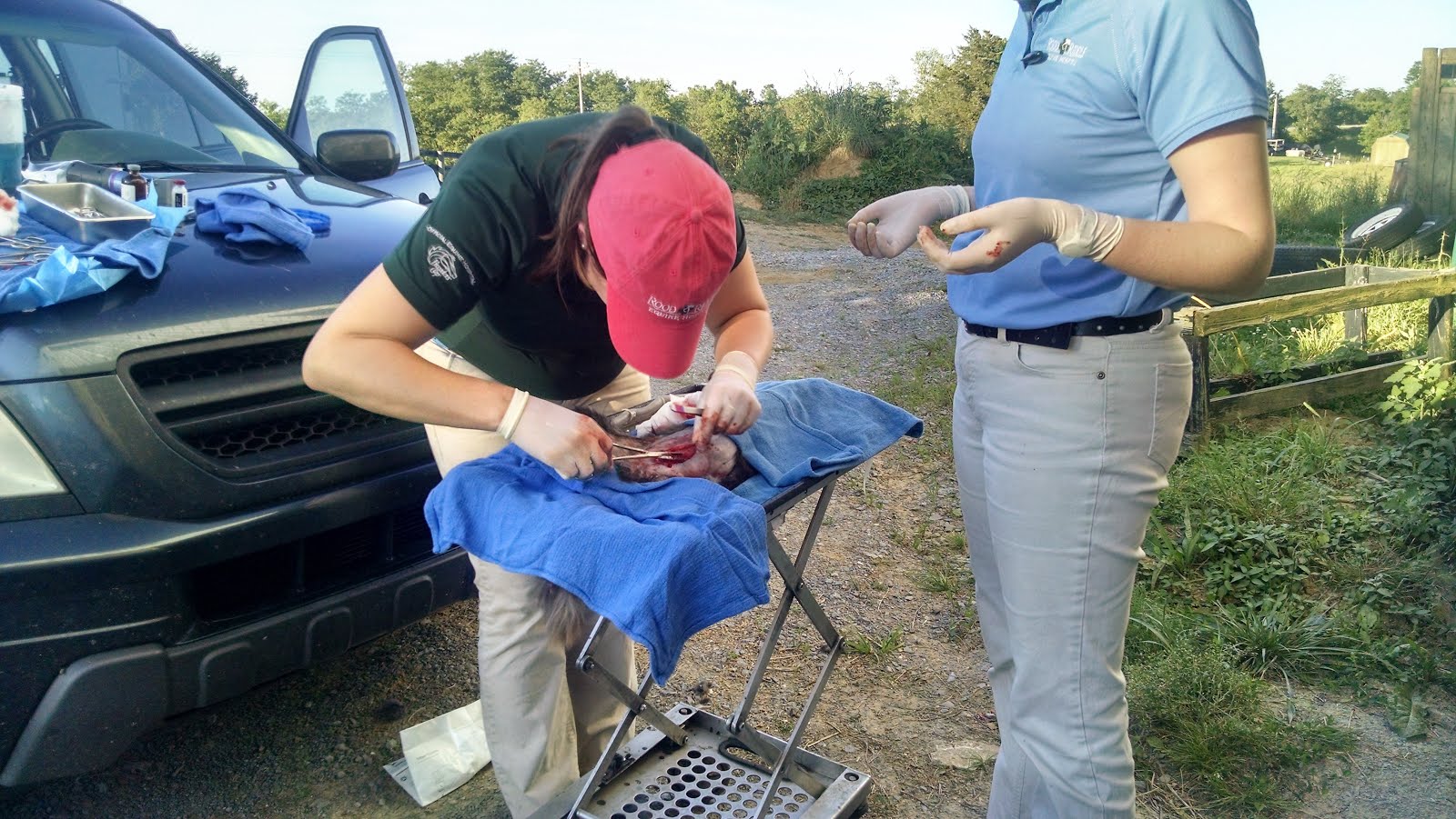
A NG tube was placed and approximately 3 gallons of fetid material was refluxed from his stomach, and he immediately perked up. He was brought into the ICU for the safety of the other horses in the clinic, and was catheterized for fluids. He received a bolus of 10 L of LRS with CMPK (due to a low calcium on bloodwork), and then was given a continuous rate of 1L per hour. He was placed on K-penicillin, banamine and gentacin. The NG tube was left indwelling, and he was refluxed about every 2 hours, resulting in less and less material until 10:0PM, then the tube was pulled at 3:00AM that night. Fluids were discontinued the next afternoon. He was ultrasounded again the next day and he showed mild improvement as his small intestine was back into a more normal position and was starting to contract and have peristaltic waves, all be it not fully yet. Though now, he began to develop a fever of unknown origin. This was handled using banamine, although he began to regulate his temperature on his own, without use of drugs. As of today (6/27/16) he has begun to develop a slight pleural pneumonia. There are some comet tails on ultrasound of his lungs and very slight fluid. Comet tail artifacts are a result of fluid accumulation in the interlobular septa of the lung. Lung radiographs were also taken and indicated only inflammation of the lungs. The horse's serum amyloid A has and is currently rising throughout his stay. There is a clear inflammatory process occurring, but it is yet to be known the exact cause of all of his problems. The pneumonia could be a result of aspiration from being refluxed, however it could also be coincidental as he did come in with slightly increase lung sounds. I am very interested to see if we can learn what his primary problem is and what is causing all of his problems. This case of enteritis, ileus, fever of unknown origin, and now pneumonia has really challenged me to think about all of the differentials and diagnostics that can be done in cases such as this. It has also given me a chance to really appreciate and learn abdominal and thoracic ultrasound. I was given the opportunity to scan him myself and was able to get some images of intestine and lung. We will see how he does and what develops over the next few days.
-Roya Oliai
Monday, June 27, 2016
Sunday, June 26, 2016
Clinton Week 2 and 3
It has been an interesting couple of weeks at CVS with variable cases.
There have been a number of horses needing work on their teeth the recently. A number of cases have involved learning how to troubleshoot. Some of the barns we have gone to have not had the best set up for the dental head stall. Many times our rope has not been long enough to reach rafters and a long line or couple of lead ropes have been borrowed to make the rope reach the halter. At another point, a mini's head was too small for the headstall and we had to figure out how to keep her in place so she wouldn't walk through the halter and end up with it around her neck. I learned that sometimes it is hard to find the correct balance with sedation to keep the horse standing but sedate enough to be worked on. This was especially true with one horse who fought against having his teeth done and tossing his head when working on the right side of his mouth, where he had many sharp points and a ramp to his molars. Another horse was brought into the clinic with a suspect tooth abcess. On dental exam, all teeth looked normal, however the second premolar on the lower left was very sensitive. Radiographs were taken and it was found there was a fracture of the second premolar. The jaw was also fractured and draining tract was noted from the jaw. The owners elected for conservative treatment and to reassess in three months.
One case was a horse with Potomic horse fever. The owner had noted in the morning that the horse seemed depressed. As the day progressed, she noted he was off feed, not drinking much and had soft feces. He had a fever over 104 F. Feces were collected for PCR and a three day course of antibiotics was perscribed. Blood work showed a leukopenia with monocytosis and lymphpenia highly suggestive of Potomic. The horse responded well to antibiotics with his temperature returning to normal the next day. The case really highlighted the debate over the efficacy of the vaccine. The horse had recieved his booster a little over a week before becoming sick. However, his fever broke quickly and he fever developed severe diarrhea.
Early one morning we had a 28 year old horse with colic. When we arrived, the mare was very distressed, bloated, and painful. Every time her handler stopped moving her, she tried to through herself to the ground. She responded to sedation and analgesia enough to perform a better exam. On rectal, there was a very tight band coursing across the pelvic inlet and a number of small abnormal lumps were felt. On ultrasound, sacculations of the ventral colon were visible in an abnormal location on the left side near the nephrospleenic space. Distended, thickened small intestine was also noted. Based on the findings, the severe pain, and the age of the horse, the owner elected for euthanasia. It was the most humane action for the mare.
Another sad case also followed up on. We visited a farm for routine work on a number of horses and to check on a foal that Dr. Trombley had been following since birth. The foal was orphaned at birth because the mare was euthanized following a utrine prolapse. Soon after, it was noted the foal was having trouble standing due to contracted tendons. Splints were placed but unfortunately the foal developed rubs from the splints that became infected. The wounds were addressed and had started to heal. However, it appears the foal had developed a septic joint after the last vet visit and the bottom of his foot rotted out, to the point where the lower bones came out. When the owner found a chunk of bone in the stall, she elected to euthanize the foal. Upon examining the chunk of bone, we discoved it was actually an intact P2. It was a very unfortunate situation. Below are pictures of P2.


It was a busy couple of weeks with some difficult cases and a lot of learning.
Equine Analysis weeks 3-6
Hey everybody!
Sorry for the delay since the last update. Since I am not in a clinic like my peers, I don't have new cases to report on each week. Right now I am still in the data collection stage so not much has changed since my last post. We are still going to Churchill Downs about three days a week and collecting data on the horses that are breezing 5/8 that day. As a reminder we perform a wind, motion, and blood test on each horse and then evaluate the data back at the office. We have data on over 30 horses now ranging from proven Grade I winners to 2 yr olds in training.
I am mostly responsible for helping to analyze the sound files collected by the wind test. I edit every breath of the sound file and make sure all of the inhales and exhales are recorded correctly. This can be very tedious as a horse breathes in and out every .2 of second when they are breezing. However, it is amazing to see the difference in breathing styles and efficiency between the great horses and those that aren't quite suited to racing. I'm also starting to be able to recognize different upper airway pathologies just by sound. I've listened to so many sound files that sometimes I hear them in my sleep! The rest of the team, the other techs and vets, have been working on analyzing the other data sets. With all of the data getting analyzed, we are finally able to start putting it all together and predicting the performance ability of each individual horse. Since some of the horses are actively racing, we also follow their racing record and see if how they run matches how we predicted they would.
Last weekend we went to Churchill Down to watch the races as some of the horses in our study were running that night. It was really fun to be on the other side of the track. We are usually working on the backside of the track and it was nice to be on the frontside for once. We went the night Gun Runner won the Matt Wynn and the crowds enthusiasm was infectious! Below are some pics from that night.
On days that I'm not analyzing data I get to travel to the other farms in the area. I've gotten to see amazing training centers and stallion barns. All of the farms are beautifully landscaped and the barns look like mansions! There are also famous horses kept at all of these farms and it is unreal to be in close contact with them. I am also planning on shadowing the track vet for a day and seeing exactly whats involved in race track medicine.
We are still having issues getting our endoscope to work but the upside is that I've gotten to practice passing the scope every time we test it out. In the mean time I've gotten to evaluate a few of the horses' airways with the track vet's scope. It definitely takes lots of practice to get used to driving and maneuvering the scope! Luckily most of these racehorses are used to being scoped and are very patient with me.
Stay tuned to see how our summer project progresses.
Sorry for the delay since the last update. Since I am not in a clinic like my peers, I don't have new cases to report on each week. Right now I am still in the data collection stage so not much has changed since my last post. We are still going to Churchill Downs about three days a week and collecting data on the horses that are breezing 5/8 that day. As a reminder we perform a wind, motion, and blood test on each horse and then evaluate the data back at the office. We have data on over 30 horses now ranging from proven Grade I winners to 2 yr olds in training.
I am mostly responsible for helping to analyze the sound files collected by the wind test. I edit every breath of the sound file and make sure all of the inhales and exhales are recorded correctly. This can be very tedious as a horse breathes in and out every .2 of second when they are breezing. However, it is amazing to see the difference in breathing styles and efficiency between the great horses and those that aren't quite suited to racing. I'm also starting to be able to recognize different upper airway pathologies just by sound. I've listened to so many sound files that sometimes I hear them in my sleep! The rest of the team, the other techs and vets, have been working on analyzing the other data sets. With all of the data getting analyzed, we are finally able to start putting it all together and predicting the performance ability of each individual horse. Since some of the horses are actively racing, we also follow their racing record and see if how they run matches how we predicted they would.
Last weekend we went to Churchill Down to watch the races as some of the horses in our study were running that night. It was really fun to be on the other side of the track. We are usually working on the backside of the track and it was nice to be on the frontside for once. We went the night Gun Runner won the Matt Wynn and the crowds enthusiasm was infectious! Below are some pics from that night.
 |
| The historic twin spires! |
 |
| In the starting gate about to take off |
 |
| Racing for home |
We are still having issues getting our endoscope to work but the upside is that I've gotten to practice passing the scope every time we test it out. In the mean time I've gotten to evaluate a few of the horses' airways with the track vet's scope. It definitely takes lots of practice to get used to driving and maneuvering the scope! Luckily most of these racehorses are used to being scoped and are very patient with me.
Stay tuned to see how our summer project progresses.
Friday, June 24, 2016
Equine Athlete Week 5!
Hey everyone, sorry for the delay in my post! The last few weeks have been filled with long days and lots of travelling, which for me means passing out as soon as we get back to the hotel. Since last time, we have traveled all over Michigan, Indiana and Ohio preparing horses for the last few regional championships of the summer and youth nationals. The diversity of the Arabian breed still continues to surprise me. All of the shows include purebred Arabian classes and half-Arab classes. The half-Arabs can be crossed with any other breed. Owners choose to breed the other half with breeds that will compliment which ever distinct discipline the horse will be performing in. A few of the different divisions that I've seen are English, country, hunter, park, side saddle, costume and western pleasure. To name a few, I've seen crosses with saddlebreds, hackneys, Dutch harness horses, and quarter horses. If you have never seen an Arabian "park" class I definitely recommend youtubing a video! In the last few weeks I've been able to practice my IV injection skills, learned and performed shock wave therapy and high intensity laser therapy on muscle, ligament, tendon and some bone injuries, flexed lots of horses for lameness exams, practiced aseptic technique, and palpated the landmarks for most joint and nerve block injections(P.S. the 2011 "Equine joint injection and regional anesthesia" book by Moyer and Schumaker has been EXTREMELY useful and I definitely recommend it to anyone interested in sport medicine). Some of the most interesting joints and areas I've seen injected so far are the poll and the shoulder, between the cervical vertebral facets and the SI region(both ultrasound guided), and injection of the navicular bursa which is performed with the help of radiographic images. I also had a few exciting firsts! At the previous show, the Buckeye Sweepstakes, I placed my first IV catheter for a horse that was shipping home and needed a little extra hydration, and during one of the routine barn visits I got to work through an entire diagnostic lameness case. This began with watching the horse lunge, flexion tests, hoof testers and palpation. The next step was then to block the suspected area. I performed my first palmar digital nerve block! Once the block set in we watched the horse lunge again. We noticed a major improvement with the block, so I then performed my first joint injection in the coffin joint! I'm currently at the Region 13 Championships in Springfield, Ohio. I will be at this show until Sunday and then I have next week off to stand up in my best friend's wedding. After next week we have two weeks of travelling to client's farms to prepare their horses for the next show, Youth Nationals in Oklahoma City. This summer has been crazy busy, but I am learning so much from Dr. Hill and Dr. O'Cull and I wouldn't have it any other way! I hope everyone is having as good of a summer as I am!
Cheers, Alex
Tuesday, June 21, 2016
Week 5 at Rood and Riddle Equine Hospital in the Ambulatory department
Taking time off for my family reunion messed my personal schedule up. I had been taking Thursdays off so that I would be able to help more on the weekends when the tech isn't working, but I didn't take the Thursday before off because I would have Sunday and Monday. Things haven't gotten back to normal yet.
Sunday the 12th was the reunion. It was good to home to see everyone and relax.The reunion was the same as always, lots of somewhat familiar faces and a few new and a ton of food! The connecting generation is my grandmother's grandparents. They have long since passed and the next generation is getting to be in questionable health. My great-grandparents are all gone now. It's getting ever more difficult for me to remember how everyone fits together on the family tree. We all ate so much more food than we really needed to.
Monday was less fun because I had to finish a few chores and leave town. I ended up running rather late and still had to run back to grab things twice. Even after that I forgot my belt. I barely made it out of the house in time to make it to my lunch date with my boyfriend. Luckily for me, he has a rather flexible lunch time. The drive back down to Lexington was uneventful.
Tuesday, I rode with Dr. Friend. We had a full truck and a full day. We started off the morning with a new foal exam and a colic exam on the new momma. We treated her for dehydration. I got to watch Dr. Friend do a bunch of chiropractic exams and adjustments on a group of yearlings. That was very interesting. We did a lameness exam on a horse that turned out to be neurologic in the hind end and also lame in the front. Together with the client, we determined that those issues plus her rough manner made her a candidate for euthanasia. We went on to a couple more stops while arrangements were made. The client didn't want to see the procedure, so we were able to practice placing catheters before the hauling company got there. Afterward, we did a couple inseminations. We went out to Dr. Friend's farm to learn/ practice hoof trimming. Unfortunately, a storm cut us off. We got caught in the downpour as we were trying to catch one of the mares to give her a shot. Then we had to go check on the colicky mare from first thing. Despite being soaking wet, it was a fun day.
Wednesday was a pretty standard repro day with Jordan. We got done very early, around 2, so I decided to finish out the afternoon (and evening) with Friend. We went to his farm again to finish what we had started the previous evening. I started a foot, but the mare saw a calf in the neighboring pasture and wouldn't let me continue. Dr. Friend had to battle her to get the hoof finished. I had already discovered that farrier work was not my cup of tea, but I wanted to learn to do it just in case I ever needed to. I was able to start on another mare and do about 3/4 of a foot before we got called out to an emergency. The call was for an unconscious foal. The barn manager and some of the workers had been watching a mare and foal run around a large paddock when, all of a sudden, the foal was not with the mare anymore. They found her unconscious next to the fence. Cranial palpation and radiographs revealed a heavily suspected crack in the cranium, most likely causing swelling on the brain. We treated for dehydration and swelling and left overnight care instructions. Not enough progress was made by morning, so they decided to euthanize the little guy. Then we went to see a club foot horse for a pre-purchase exam. We ultrasounded the tendons and discussed the added risk of using the horse. Our final stop for the night was at a nice barn to see a horse with bilaterally swollen hocks. We took synovial fluid samples from both and flushed the one that he was lame on. Wednesday turned out to be a cool for teaching but unfortunate for owners day.
Thursday was a good day. We found a bunch of embryos and fetuses on pregnancy and heart beat checks. We took some pastern radiographs and floated some teeth. We got a call from a client whose horse likes to fight with his friend. He has a bunch of old bite wounds all down his neck. One of them caused an abscess which had ruptured out the other side. It was a very delicate situation because it had opened in the jugular groove just superficial to the jugular vein. We lavaged the area, and Jordan very carefully placed a drain tube to help it heal correctly.
Friday was a late start. It was wonderful to get to sleep until a normal time for people to be getting up for work. It was very strange though for it to be so light outside and so much traffic. We had a typical repro morning with lots of palpation, pregnancy checks, and lavages. In the afternoon, we went out to vaccinate a pony and euthanize a goat. That was an interesting experience because we had to dilute most of the drugs we used. Our final stop of the day was a hoof sole puncture. The horse had managed to partially remove her new fancy shoe and step on the clip on the front. Since the puncture was so thin, we weren't able to lavage or do much of that. We helped get the foot into a CleanTrax soak and instructed the client to pack and wrap the foot to keep the sole clean until it healed. They were already doing most of that from the farrier's advice. We were able to add some Bute and antibiotics to the mix.
Saturday was so slow that I was given the day off. It seems that people (and horses) have started trying to make weekends a thing again, especially the holiday weekends. I had a fairly productive day of pre-treating some laundry, grocery shopping, and showering. In the evening, I discovered that I was also being given Sunday off, so I decided to run home to surprise my boyfriend for Father's Day.
Weeks 5 and 6 as a RREH Surgery Tech Student
Weeks 5 and 6 marked another transition for me. I’ve started
to shift from a need for help acting as a tech for most surgeries to a greater
independence in the surgery room. The more I learn about routines and where
things are located, the more comfortable and relaxed I can be during a surgery.
Screw placements to correct for angular limb deformities (ALDs) are the surgery
in which I’m most comfortable. These surgeries are generally simpler to tech
because once you have helped the clinician “set up” for the surgery (connect
the drill, drape the patient) you have more time to set up for the next surgery;
the room should be ready for the next technician to bring a patient in when you
are done. Taking radiographs is a guarantee with screw placement regardless of
the clinician involved, which means that you can be ready for them. Radiographs
are taken to confirm that the screws are placed appropriately to correct for
the ALD. I have yet to see a screw placement that had to be adjusted!
Arthroscopies (joint surgeries to clean out bone fragments and cartilage
buildup) are a little more complicated however. Because of the high number of
arthroscopies each day and the high cost of the instruments (it takes a camera,
and a light among other things), the instruments are sterilized between
surgeries in a very strong chemical that you have to be careful working with.
As technician you are responsible for sterilizing the instruments for the next
arthroscopy and assisting in removing the instruments from the chemicals for
your surgery. Additionally, fluids are run through the joint that is being scoped
in order to provide a clean camera view for the surgeon while simultaneously
acting as a joint flush. The technician is responsible for hanging and hooking
up fluids to the fluid pump, hooking up the camera and light all the while
keeping up with the surgeon’s needs. Needless to say, it takes a bit more
finesse and experience to tech an arthroscopy as well as I can tech a screw
placement. I also teched my first tie forward at the end of week 6, and a
couple of closed castrations. A tie forward is performed to suture the larynx
in such a way that the epiglottis sits closer to the pharyngeal opening so as
to correct for dorsal displacement of the soft palate (over the epiglottis), a
problem that can hinder performance horses by making their airway smaller. A
tie forward is widely considered to be the most beneficial and successful
surgical option for a displaced soft palate in a horse. Teching a tie forward
is most involved in the preparation for the surgery. Clipping around the neck
and jaw area of the horse is a more sensitive and particular procedure. Prepping
the surgical site involves covering the horse’s eyes so that the cleaning
solution doesn’t drip into them and damage the cornea. Finally, closed
castrations are a more simple surgery to tech, as there are no fluids to run or
radiographs to take (etc.). Prep the surgery site (with a less intense chemical
scrub), adjust the lighting, keep some extra suture on hand and you’re good to
go! Add a few intermittently placed colic surgeries to the list and you’ll have
a good idea of how the past two weeks have gone.
A couple of the interesting sights these past two weeks
include a miniature horse colic that had abnormally formed intestine with
unusual (and sometimes lacking) mesenteric attachments, and a laceration that
had devolved into something much more aggressive-looking (which ended up
looking much better after a few days of treatment).


The laceration!
Monday, June 20, 2016
Weeks 4 and 5 at Littleton
Hello from Littleton!
It has been a very busy past couple weeks. The Summer Show Series is in full swing at the Colorado Horse Park, I spent my first weekend of 7 out there this past weekend, and we certainly had a full schedule! Littleton Equine is the official show vet there, so someone is basically always around. We saw a lot of the typical health certs, PPE's, joint injections, a lot of lameness exams and thus following with a million and one rads..! At least, it feels like it :D
I was out with one of the new interns, Dr. Katy Mayhew, and Dr. French was our main staff vet on hand. Dr. French does both acupuncture and chiropractic as well, so there were a lot of adjustments and needles, which was great because I have found a really strong interest in both of those areas.
There was a horse that developed a fever and some bilateral nasal discharge, so it was important to keep monitoring him closely. This also led to discussions and actions about creating some isolation areas out there at the park for when situations like this arise. So that's exciting and in the works to finally have a designated iso area.
There was also the fair share of fun as well, we had a couple opportunities to watch the Grand Prix show jumping Saturday night, where some of Littleton's clients got 1st and 3rd. Sunday was the Hunter and Jumper Derby's and thankfully no injuries, but they certainly push those horses hard. It was great to see some fantastic athletes at work.
As for my other days, for the most point I was back in the hospital, either in ICU or surgery days. We had a pretty busy week in ICU, some colic recoveries, one with a potential abdominal abscess that we were trying to get and stay in front of, another snake bite, and some mares with their foals, for various reasons. Thankfully it was a pretty clean bill of health for most of them.
On surgery, there was also quite the variation. Along with the normal arthroscopes, we had another arthrodesis, except this one was in the hock. Dr. Devine just went in to the TMT and DIT joints and removed some of the cartilage remaining, and predicts that within 4-6 months, ideally, the joint will be fused. He tells clients to give it a year though, just to really make sure. I was also surprised with the drastic differences within joints for arthrodesis. How much range of motion, the location, etc all of those factors that play into the intensity of the procedure.
We also had a mare come in with a broken mandible and numerous scratches and lacerations. The owners were unsure of what had happened, unfortunately, so it was kind of a mystery. Dr. Devine used 3 cerclage wires to realign and secure the mandible together. He said that they could come out after about 6-8 weeks, do some rechecks to make sure, but if there is the contact within the bone, then it should be successful. We were all very curious as to what had happened!


One interesting thing that I learned the other week that is somewhat unique to Colorado/high altitude areas is the use of isoxsuprine in the treatment of some navicular horses. The drug is mostly a cardiac drug for humans, but after speaking with Dr. Beeman out here, he says that it seems to have some affinity for horses as well. The problem with navicular cases here, is that at a higher altitude, liquids and gases expand, thus increasing pressures. This can cause more pain in a constricted area like the foot. Isoxsuprine has been shown to increase the venous return (not necessarily the volume), and acts as a vasodilator, so when given here, it has a relieving effect. Dr. Beeman also said that many people in lower elevations don't see the same results, due to differences in pressures. I found this really interesting! The altitude here and even higher into the mountains presents some really unique challenges to our animals and it's been intriguing learning some of these differences from the sea level of Michigan.
Again, as I said, this summer has not been without fun..! Dr. Devine, our surgeon, actually front lines a country band, so I was able to go up to a small town called Evergreen where Dustin Devine and the Real Deal had a gig at the Little Bear bar! It was really fun to see some people outside of the hospital, and he showed off many more sides of talent! Looking forward to another exciting week here, hope everyone's summers are going great too!
-Taylor Alton
It has been a very busy past couple weeks. The Summer Show Series is in full swing at the Colorado Horse Park, I spent my first weekend of 7 out there this past weekend, and we certainly had a full schedule! Littleton Equine is the official show vet there, so someone is basically always around. We saw a lot of the typical health certs, PPE's, joint injections, a lot of lameness exams and thus following with a million and one rads..! At least, it feels like it :D
I was out with one of the new interns, Dr. Katy Mayhew, and Dr. French was our main staff vet on hand. Dr. French does both acupuncture and chiropractic as well, so there were a lot of adjustments and needles, which was great because I have found a really strong interest in both of those areas.
There was a horse that developed a fever and some bilateral nasal discharge, so it was important to keep monitoring him closely. This also led to discussions and actions about creating some isolation areas out there at the park for when situations like this arise. So that's exciting and in the works to finally have a designated iso area.
There was also the fair share of fun as well, we had a couple opportunities to watch the Grand Prix show jumping Saturday night, where some of Littleton's clients got 1st and 3rd. Sunday was the Hunter and Jumper Derby's and thankfully no injuries, but they certainly push those horses hard. It was great to see some fantastic athletes at work.
As for my other days, for the most point I was back in the hospital, either in ICU or surgery days. We had a pretty busy week in ICU, some colic recoveries, one with a potential abdominal abscess that we were trying to get and stay in front of, another snake bite, and some mares with their foals, for various reasons. Thankfully it was a pretty clean bill of health for most of them.
On surgery, there was also quite the variation. Along with the normal arthroscopes, we had another arthrodesis, except this one was in the hock. Dr. Devine just went in to the TMT and DIT joints and removed some of the cartilage remaining, and predicts that within 4-6 months, ideally, the joint will be fused. He tells clients to give it a year though, just to really make sure. I was also surprised with the drastic differences within joints for arthrodesis. How much range of motion, the location, etc all of those factors that play into the intensity of the procedure.
We also had a mare come in with a broken mandible and numerous scratches and lacerations. The owners were unsure of what had happened, unfortunately, so it was kind of a mystery. Dr. Devine used 3 cerclage wires to realign and secure the mandible together. He said that they could come out after about 6-8 weeks, do some rechecks to make sure, but if there is the contact within the bone, then it should be successful. We were all very curious as to what had happened!
One interesting thing that I learned the other week that is somewhat unique to Colorado/high altitude areas is the use of isoxsuprine in the treatment of some navicular horses. The drug is mostly a cardiac drug for humans, but after speaking with Dr. Beeman out here, he says that it seems to have some affinity for horses as well. The problem with navicular cases here, is that at a higher altitude, liquids and gases expand, thus increasing pressures. This can cause more pain in a constricted area like the foot. Isoxsuprine has been shown to increase the venous return (not necessarily the volume), and acts as a vasodilator, so when given here, it has a relieving effect. Dr. Beeman also said that many people in lower elevations don't see the same results, due to differences in pressures. I found this really interesting! The altitude here and even higher into the mountains presents some really unique challenges to our animals and it's been intriguing learning some of these differences from the sea level of Michigan.
Again, as I said, this summer has not been without fun..! Dr. Devine, our surgeon, actually front lines a country band, so I was able to go up to a small town called Evergreen where Dustin Devine and the Real Deal had a gig at the Little Bear bar! It was really fun to see some people outside of the hospital, and he showed off many more sides of talent! Looking forward to another exciting week here, hope everyone's summers are going great too!
-Taylor Alton
Saturday, June 18, 2016
Week 4 with RREH Ambulatory
The last 2 weeks have gone by so fast! We've done a lot and seen a couple of really interesting cases. Some of the things have been cool teaching experiences that weren't so good for the owners. I'm still loving every minute of it!
Sunday the 5th was the first rather slow day we had. I went out with Dr. Friend, but we didn't have much on the books. I picked up the clinic van for him, so he could take his truck in to be looked at on Monday. We went to one farm and did a bunch of uterine flushes, then he brought me back to the clinic. I helped him restock the truck before he left for one other farm. I had a relaxing day catching up on some Netflix and eating a full 3 meals.
Monday I was with Jordan. She had been out late with an emergency that turned out to be NSAID toxicity. The lesson there was to pay close attention to the dosing on your medications. Just because 1 gram was 3 spins on one container doesn't mean it always will be. Monday was a pretty standard day with palpations and lavages. I got to hold the plate for some foot radiographs and saw how to upload images.
Tuesday, we started off the day with a couple pregnancy checks and treating a pair of foals with puncture wounds. We took some radiographs of a yearling. We finally had time to sit down to a nice lunch with Dr. Friend and his group. We all went together to cover for another vet who was on vacation. We got a call for a violent colic. Many drugs and lots of effort later, we got the mare on the trailer to the clinic. Later we learned she had a torsion just behind the cecum. She made it through surgery but went downhill and was euthanized the following day. After the emergency, we saw a heel bulb abscess. The past 2 days made me realize to never worry about filling up your day. Things will always come up.
Wednesday was an early morning because we had a few things to get done before we started our shift at the Horse Park. We ended up having to do 2 new foal exams and run plasma to each of them at our second stop. That made us a touch late to the Horse Park, but no one seemed to mind. The only thing that came up at the Horse Park was one case of hives. After that we inseminated a mare, found a couple beautiful pregnancies, chased a baby around a field, microchipped said baby. Jordan stitched up a torn eyelid and I met the eldest Friend child.Our final stop took us south of the river. We ended up spaying 2 cats on the cart.

 The spays were very fun for me because I got to find half of the uterus, place a few sutures, and tie a few sutures. It was interesting to see how Jordan was able to make do with the things we had to help control the cat population on this farm.Thursday, we started the day replacing leg wraps on a foal fighting contracted tendons. Then we sat around the Horse Park for a couple hours. The rest of the day was pretty standard. I did a uterine flush on my own. Jordan dropped me off around 7 and I went to one of our big farms to watch a foaling. The night manager walked me through the process and then we waited. Another lady came to help since the night manager had been watching this mare for a couple days with very little sleep. I kept falling asleep in the chair waiting for something to happen.

I got lots of kitty cuddles from one of the barn cats.Finally around 4:30 Friday morning, the mare's water broke. I was amazed at how quickly she was able to get the foal on the ground. We checked on the other mare that was showing some of the early signs of foaling and found that her water had broken as well. By 5 am, we had 2 new babies to handle. I got the day off because I didn't get home until 7:30. I slept until 3:30 pm and rested and Netflixed the rest of the day,
Saturday, I was back with Dr. Friend. We did a lot of lavaging. I got to do the 2 post-foaling lavages on the mares that foaled last week. That was a very interesting experience. We took out some leg sutures and did a lameness exam. I got to do my first insemination with fresh semen. I took some things back to the clinic and headed home for a family reunion while Dr. Friend met Jordan to do a hernia banding and start monitoring for frozen semen timing.
I made it home in time to visit my old roommate at her puppy birthday party. I had a great week and enjoyed being home for a bit.
Sunday the 5th was the first rather slow day we had. I went out with Dr. Friend, but we didn't have much on the books. I picked up the clinic van for him, so he could take his truck in to be looked at on Monday. We went to one farm and did a bunch of uterine flushes, then he brought me back to the clinic. I helped him restock the truck before he left for one other farm. I had a relaxing day catching up on some Netflix and eating a full 3 meals.
Monday I was with Jordan. She had been out late with an emergency that turned out to be NSAID toxicity. The lesson there was to pay close attention to the dosing on your medications. Just because 1 gram was 3 spins on one container doesn't mean it always will be. Monday was a pretty standard day with palpations and lavages. I got to hold the plate for some foot radiographs and saw how to upload images.
Tuesday, we started off the day with a couple pregnancy checks and treating a pair of foals with puncture wounds. We took some radiographs of a yearling. We finally had time to sit down to a nice lunch with Dr. Friend and his group. We all went together to cover for another vet who was on vacation. We got a call for a violent colic. Many drugs and lots of effort later, we got the mare on the trailer to the clinic. Later we learned she had a torsion just behind the cecum. She made it through surgery but went downhill and was euthanized the following day. After the emergency, we saw a heel bulb abscess. The past 2 days made me realize to never worry about filling up your day. Things will always come up.
Wednesday was an early morning because we had a few things to get done before we started our shift at the Horse Park. We ended up having to do 2 new foal exams and run plasma to each of them at our second stop. That made us a touch late to the Horse Park, but no one seemed to mind. The only thing that came up at the Horse Park was one case of hives. After that we inseminated a mare, found a couple beautiful pregnancies, chased a baby around a field, microchipped said baby. Jordan stitched up a torn eyelid and I met the eldest Friend child.Our final stop took us south of the river. We ended up spaying 2 cats on the cart.

 The spays were very fun for me because I got to find half of the uterus, place a few sutures, and tie a few sutures. It was interesting to see how Jordan was able to make do with the things we had to help control the cat population on this farm.Thursday, we started the day replacing leg wraps on a foal fighting contracted tendons. Then we sat around the Horse Park for a couple hours. The rest of the day was pretty standard. I did a uterine flush on my own. Jordan dropped me off around 7 and I went to one of our big farms to watch a foaling. The night manager walked me through the process and then we waited. Another lady came to help since the night manager had been watching this mare for a couple days with very little sleep. I kept falling asleep in the chair waiting for something to happen.
I got lots of kitty cuddles from one of the barn cats.Finally around 4:30 Friday morning, the mare's water broke. I was amazed at how quickly she was able to get the foal on the ground. We checked on the other mare that was showing some of the early signs of foaling and found that her water had broken as well. By 5 am, we had 2 new babies to handle. I got the day off because I didn't get home until 7:30. I slept until 3:30 pm and rested and Netflixed the rest of the day,
Saturday, I was back with Dr. Friend. We did a lot of lavaging. I got to do the 2 post-foaling lavages on the mares that foaled last week. That was a very interesting experience. We took out some leg sutures and did a lameness exam. I got to do my first insemination with fresh semen. I took some things back to the clinic and headed home for a family reunion while Dr. Friend met Jordan to do a hernia banding and start monitoring for frozen semen timing.
I made it home in time to visit my old roommate at her puppy birthday party. I had a great week and enjoyed being home for a bit.
A Rather Warm Week At Oakridge Equine
It's been quite hot here in Oklahoma this week! With temperatures in the triple digits, I have never been more grateful for air conditioning!
After our busy week of colics last week, this week seemed pretty quiet. We only had a handful of emergencies including a couple colics and a laceration, only two that needed to go to surgery though.
Throughout the week we had pretty routine cases, including many lameness exams, pre purchases, MRIs, EPM work ups, joint injections and arthroscopies.
Some of the more interesting cases were one of the MRIs was of the brain so it was pretty cool to read that report. The horse had been having episodes of seizures and unpredictable behavior, so with a history of a possible head injury, they decided to see if there was any indication of brain damage. I never had a chance to see the final report, but the preliminary findings didn't show any indication of injury.
We also saw several horses this week for patellar ligament splitting, where small incisions are made into the ligament to cause thickening and scaring down of the ligament to help with horses who have problems with locking their stifles. It's a pretty quick standing procedure.
Another case we saw was a mare with a sequestrum on her pelvis. Dr Lamb was able to remove this standing and it was a pretty cool procedure to watch. It was a very large sequestrum!
Another procedure I've never seen before is cutting the deep digital flexor tendon. This is done to help reduce rotation of p3 to keep the horse more comfortable. Although not an ideal situation, this mare was expected to make a good recovery to hang out in a pasture for her retirement years.
For a good way to end the week, the zoo brought a gorilla down for an MRI on Friday.
That's it for now! Hopefully by next update it will have cooled off a bit here!
Friday, June 17, 2016
Weeks 4-6 at Stevens Equine
Well, horse show season is officially in full swing! The past few weeks have flown by and have been filled with long days of a lot of lame horses and some interesting cases. Other than today, the past several days have been very hot and humid so we have had to administer fluids, electrolytes, and some B vitamins to a few horses to help hydrate them and perk them up a bit. Other than that, there has been the usual mix of lameness examinations that are often followed up with either more in-depth diagnostics of ultrasonography or radiographs and intra-articular injections or other treatments.
As usual for horses, there have been several cases lately where horses have gotten lacerations in creative ways. One horse got its leg caught in an arena fence, one caught its lower eyelid on something in the stall, and two got their leg entrapped in the stall door bars. I am always amazed to see how they find unique ways to maim themselves, but fortunately all of those were able to be sutured and/or bandaged and treated with anti-inflammatories and antibiotics so are recovering well.
Although I have bought and sold horses several times before, I was able to participate in my first pre-purchase exam the other day. It was interesting helping to assess the condition of several of the horse’s systems and to evaluate its health and potential suitability as an eventing horse. This horse was an ex-racehorse, and it was very interesting listening to Dr. Stevens look through its racing record and explain the different components of it and their potential implications to the interested buyer. It made a lot of sense, and is not something that I would have thought to pore through other than out of sheer interest.
There have been a few horses that have been afflicted with laminitis over the past few weeks, and it has been neat learning about that and its correlation with Equine Metabolic Syndrome. Dr. Stevens assigned me to read the ACVIM Consensus Statement on the condition, so I learned a tremendous amount about both EMS and PPID (Pituitary Pars Intermedia Dysfunction). Both are conditions that I have always been interested in yet had not read about in-depth, so it was great learning about the diagnosis, treatment, management, predilections, etc. of them. I highly suggest reading the ACVIM paper on it if anyone is interested, because it was full of great information and was a very user-friendly report.
Lastly, I was able to watch a very interesting procedure the other day. It is called intravenous regional limb perfusion, and was performed on an older horse that has had ongoing lameness issues for a while that had been unresponsive to other treatments. We did radiographs of his distal forelimbs a few weeks prior to this particular procedure, and noted an area of abnormal radiolucency on his right front distal cannon bone that was a degenerative bone lesion that Dr. Stevens thought may respond to treatment with Tildren. Tildren is a first generation bisphosphonate that is often used to treat orthopedic conditions of the distal limb in horses. In order to use a lower overall dose and to deliver a higher amount of the drug to the site of the lesion, he used intravenous regional limb perfusion as well as administration via an intravenous jugular catheter for systemic application.
Basically what was done was that the horse was given a low four-point block to prevent any uncomfortable sensation in his lower limb during treatment then an Esmarch bandage was applied just below the carpus. The Tildren was subsequently injected into the limb via a butterfly catheter into the palmar digital vein then the site of venipuncture was covered with a temporary compression wrap and we waited for 30 minutes before removing the tourniquet. That allowed sufficient time for the drug to leave the vasculature and enter the tissues. It was really interesting to watch, and I learned that it is a procedure that is also commonly done to deliver antibiotics at a high concentration to distal limb lacerations or septic joints.
Again, I hope that everyone is having a great summer and is enjoying their hands-on learning!
Wednesday, June 15, 2016
Weeks 4-5 Cleveland Equine
One very interesting case that came into the clinic was a 3 year old Thoroughbred gelding. He was presented to us with a 30 day history of pleural pneumonia. He was being treated off and on with antibiotics--compounded chloramphenicol, but was not improving.
We ultrasounded his lungs, and found consolidation, fluid, and fibrin.

Cranial is to the left of the US, and Caudal to the right. The hyperechoic triangular portion on the left of the image is the tip of the left lung. Fluid surrounds it, with clumps of fibrin along the diaphragm. The liver can be seen as the hypoechoic wedge on the right of the image.
He was hospitalized in our ICU, and a bilateral thoracentesis was performed. Approximately 2-3 gallons of pleural fluid was drained off. The tubes were sutured and bandaged in place to allow for further drainage at later times during his stay. A three way stop-cock was placed at the ends of the tubes to close them, in order to prevent a pneumothorax.
He was also being treated with systemic antibiotics until culture and sensitivity results were obtained based on the pleural fluid. Upon admission blood work was also done on him. His Serum Amyloid A (SAA) was very high, this is an acute phase protein that indicates inflammation- it has a shorter half life than fibrin and can therefore be monitored daily to see if any improvements are being made. Globulins were high (antibodies trying to fight infection). Albumin was low (because it is leaking out of the vasculature into pleural space). Slight decrease in hematocrit (anemia of chronic infection), and also decreased lymphocytes (due to infection).
Unfortunately after about a 48 hour stay he began to deteriorate quickly. His breathing became extremely labored, and his mucus membranes began to look toxic. Another ultrasound was done at this time and portions of the lung were not moving at all. At that time the owner made the decision to euthanize. A basic necropsy of his chest cavity was done, and it was found that portions of his lung lobes had formed adhesions to the thorax wall, and other portions showed signs of necrosis.
As a part of my time at CEC I check on our inpatients, after only a couple of nights this horse became a favorite. It was sad to see him go, but harder to watch him suffer. I am glad to have been able to learn from his case and help as I could.
-Roya Oliai
Subscribe to:
Posts (Atom)