Greetings from Rood and Riddle!
These past two weeks have been busy busy in the surgery department! 90% of the surgeries seen here are often transphyseal bridging or arthroscopies. Transphyseal bridging is done on horses that are one year old or younger to correct uneven growth of a foot and usually involves inserting a staple or series of screws and wires on the knee or fetlock to inhibit growth on one side of the joint, giving the other side a chance to catch up. Once a limb has corrected, the staple is removed. If the implants are not removed, the angular deformity will overcorrect, and the leg will acquire a deformity in the opposite direction.
Arthroscopic surgery is minimally invasive surgery in which an endoscope attached to a camera is inserted into the joint via a small incision. The surgeon can then view the inside of the joint on a video monitor. Because the joint does not have to be completely opened, the recovery time is usually significantly reduced, and the success rate may be greater due to a decrease in trauma to the connective tissues around the joint. In cases of OCD or chip fractures (which is what we see almost everyday here!), the horse is placed under general anesthesia, the affected joint is prepared for surgery, and two small incisions are made in the skin overlying the joint. One incision allows placement of the arthroscope in the joint so the surgeon can visualize the lesion, and the other incision allows the instruments to be placed in the joint to allow chip fracture removal or debridement of an OD lesion.
On top of our usual days, there is always a special case in the mix which keeps things interesting! One interesting surgery we did was a cervical spinal cord surgery. This surgery is done on horses exhibiting Wobbler Syndrome, which refers to a number of disease states in the horse. The most common is termed cervical vertebral malformation and is characterized by malformation or compression of the spinal cord which leads to spasticity, ataxia, and incoordination.These symptoms are caused by damage to or compression on the spinal cord. The surgical technique involves drilling a hole between the affected vertebral bodies from underneath the neck and inserting a stainless steel prosthesis called a “Bagby Basket,” which fuses and immobilizes the vertebrae.
 Dr. Woodie performing the basket placement surgery
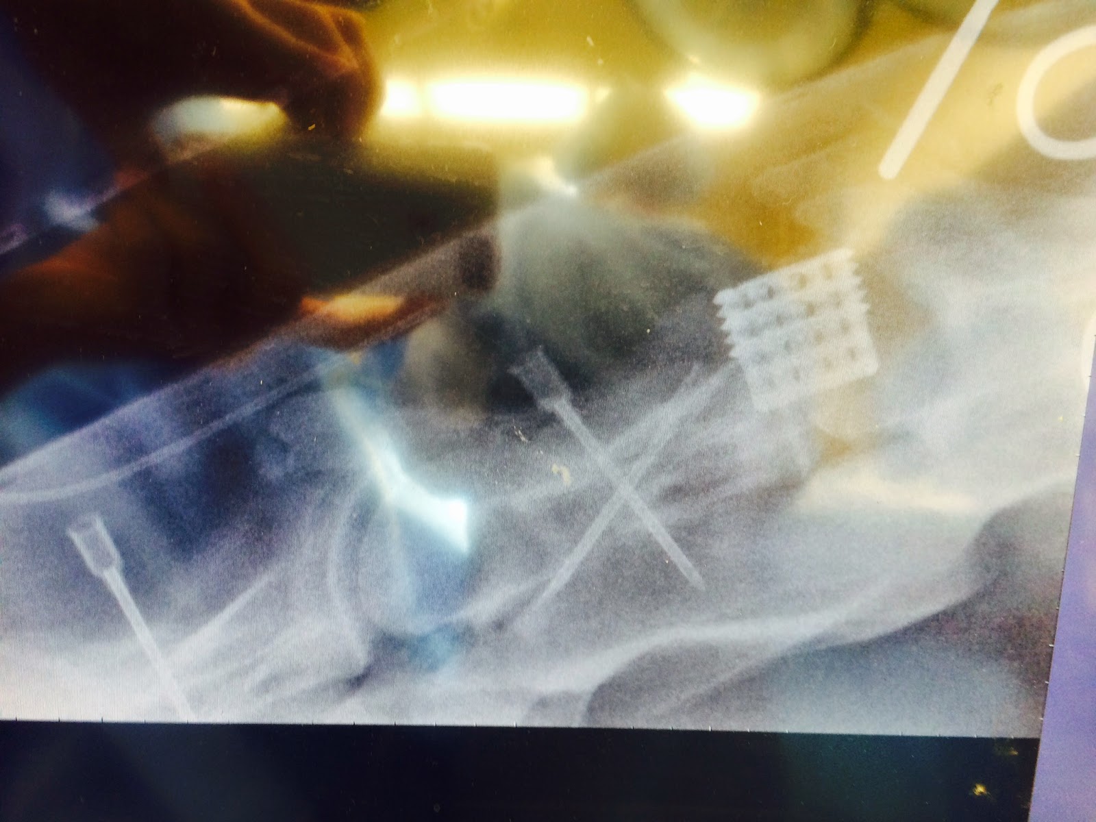 Basket implant on a radiograph of the spinal cord
Another interesting case this week was a mare brought in for an ovariectomy, which is a surgery that involves the removal of a diseased ovary. This is a commonly done procedure, and can be done on the farm. However this was a special case, as this ovary came out to be about the size of a bowling ball on ultrasound! Dr. Woodie had to remove about 1 liter of fluid out of the ovary before we could even think about trying to remove it out of the body.
 This image represents a normal ovary, with the one on the left being during non-breeding season, and on the right during breeding season.
 This is the ovary we removed, clearly diseased, and much larger.
Thanks for reading our first post from Rood and Riddle!!!!
-Lisa Reznik
These past two weeks have been busy busy in the surgery department! 90% of the surgeries seen here are often transphyseal bridging or arthroscopies. Transphyseal bridging is done on horses that are one year old or younger to correct uneven growth of a foot and usually involves inserting a staple or series of screws and wires on the knee or fetlock to inhibit growth on one side of the joint, giving the other side a chance to catch up. Once a limb has corrected, the staple is removed. If the implants are not removed, the angular deformity will overcorrect, and the leg will acquire a deformity in the opposite direction.
No comments:
Post a Comment