It is so hard to believe that I will leave Brown Equine Hospital in less than a week. This summer has flown by and I have learned so much in the short time I have spent here. Like the summer, this past week sped by and we had a full caseload of horses to be scoped.
The most invasive procedure we performed with the scope was a ventriculocordectomy. A ventriculocordectomy is the removal of the laryngeal ventricles and vocal cords to enlarge the airway. During this procedure, the scope is passed through the nose to the level of the epiglottis and gives the veterinarian a clear view of the larynx as he works. The ventricles and vocal cords can then be removed through an incision in the throatlatch area. In our first case, the patient had partial paralysis of the left recurrent laryngeal nerve, which prevented him from fully opening his left arytenoid cartilage and vocal cord. Dr. Brown is confident that the ventriculocordectomy will allow him to perform at a high athletic level despite the partial paralysis.
We suspected that a second horse presenting with exercise intolerance also had laryngeal nerve paralysis. This gelding had an even more pronounced paralysis in both the left and right arytenoid cartilages and the owner was already considering a ventriculocordectomy. Upon close examination of the horse as a whole, however, severe facial muscle atrophy on the right side of the face was also observed. Dr. Brown diagnosed the horse with Equine Protozoal Myeloencephalitis (EPM), a common neurological disease. Instead of staying for surgery, the gelding went home to be treated with anti-protozoal drugs and a vitamin E supplement.
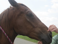

Minor muscle atrophy of the left side of the face compared to advanced muscule atrophy of the right ride
We used a gastroscope to go up the nose, under the epiglottis, down the esophagus, and into the stomach of another patient to check for gastric ulcers. Surprisingly, once we entered the stomach, we found no ulcers. Instead we found clusters of bot fly eggs (truly disgusting.) Our final scope case was the guttural pouch fungal infection that we have been treating topically through the scope for the past three weeks. We are now able to tear pieces off of the fungal plaque and inject the topical treatment directly into it. Below is a series of pictures documenting our progress.


The plaque upon presentation, during a lavage with dilute betadine, after two weeks of treatment


Topical treatment with an anti-fungal suspension, after three weeks of topical treatment
Despite being busy, we still found some time to have fun. One of Dr. Brown’s heavy weight pulling horses, Tank, has been staying at the clinic so Dr. Brown can work with him. The externs and I go along for the ride when we can to add weight to the sled; we have a grand time trying to keep our balance on the moving pallet. Dr. Brown even let me try my hand driving Tank. It is never a dull day around here!


No comments:
Post a Comment